
Accessory gland anatomy · Fertility · Prostate-cancer staging
What Are the Seminal Vesicles? Anatomy, Seminal Fluid and Ejaculatory Function
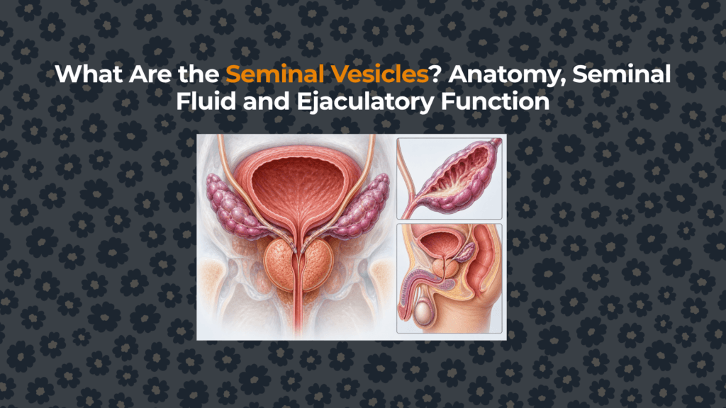
The seminal vesicles are paired glands tucked behind the bladder and above the prostate. Despite their name, they do not store sperm. Their highly folded lining produces most of the fluid in a typical ejaculate, including fructose and semenogelin, and their muscular walls contract during emission.
Where are the seminal vesicles located?
The glands lie in the pelvis between the posterior bladder wall and the rectum, superior and posterior to the prostate. Their lower ends converge toward the prostate base. This placement makes them adjacent to—yet anatomically distinct from—the prostate described in the bladder–prostate–rectum relationship guide.
Each gland is usually elongated, lobulated and obliquely oriented. The ampulla of the vas deferens sits medially. Inferiorly, the seminal-vesicle duct unites with the corresponding vas deferens to make an ejaculatory duct, which then travels through prostate tissue.
What do seminal vesicles do?
Their secretory epithelium creates an alkaline, viscous fluid rich in carbohydrates, proteins, ions and signaling molecules. Fructose is a familiar marker of seminal-vesicle secretion, but the fluid is not simply “sperm food.” Semenogelin proteins help form the temporary seminal coagulum after ejaculation; bicarbonate contributes buffering; prostaglandins and other molecules influence sperm and reproductive-tract interactions.
Prostate fluid supplies a different chemical mixture, including PSA, citrate and zinc. PSA later cleaves semenogelin and helps semen liquefy. The glands therefore work as a coordinated system, explained more fully in the prostate function and seminal-fluid article.
How does fluid reach the urethra?
Sperm travels through the vas deferens
Sperm produced in the testes and matured in the epididymis is propelled toward the pelvis during emission.
The seminal vesicles contract
Sympathetic autonomic signaling coordinates contraction of smooth muscle in the ducts and gland walls, adding seminal-vesicle fluid.
Paired ejaculatory ducts form
Each seminal-vesicle duct joins its vas deferens. The two ejaculatory ducts pass through the prostate.
Contents enter the prostatic urethra
The ducts open near the seminal colliculus in the prostatic urethra, where prostate secretions also join before expulsion.
Seminal vesicle vs prostate: what is the difference?
| Feature | Seminal vesicles | Prostate |
|---|---|---|
| Number and form | Two elongated, coiled glands | One compact gland organized into zones |
| Position | Behind bladder, above prostate | Below bladder, around proximal urethra |
| Duct pathway | Duct joins vas deferens to form ejaculatory duct | Numerous small ducts open directly into prostatic urethra |
| Typical fluid share | About 50–65% | About 20–30% |
| Characteristic products | Fructose, semenogelin, prostaglandins, bicarbonate | PSA, citrate, zinc and proteolytic enzymes |
| Embryological origin | Mesonephric (Wolffian) duct outgrowth | Urogenital sinus epithelium |
Can a person live without seminal vesicles?
Yes. Radical prostatectomy for prostate cancer usually removes the prostate and seminal vesicles together. This does not remove the testes or eliminate testosterone, orgasmic sensation or the ability to produce sperm in the testes, but it eliminates ejaculation of semen and causes infertility through intercourse. Erectile and orgasm outcomes depend on nerves, vessels, treatment details and individual health.
Congenital absence can also occur. Bilateral seminal-vesicle agenesis is often associated with absent vas deferens and CFTR-related disease; unilateral absence may accompany an ipsilateral kidney or duct abnormality. Such patterns reflect their shared embryological origin rather than damage caused by the prostate.
What conditions affect the seminal vesicles?
| Condition | Possible clues | How it is evaluated |
|---|---|---|
| Inflammation or infection | Pelvic pain, painful ejaculation, urinary symptoms, blood in semen | History, examination, urine/semen testing when appropriate, targeted imaging |
| Cyst or duct obstruction | Low-volume ejaculate, infertility, discomfort or incidental imaging finding | Semen analysis, ultrasound or MRI; evaluate associated duct/kidney anatomy |
| Stones or hemorrhage | Hematospermia, pain or imaging signal changes | Ultrasound, CT or MRI chosen for the clinical question |
| Prostate-cancer invasion | Usually identified by staging MRI or prostatectomy pathology | Integrated cancer staging; pathological invasion is pT3b |
| Primary seminal-vesicle tumor | Very rare; may present with bleeding, pain or obstruction | Imaging plus histopathology and exclusion of another primary site |
Why does seminal-vesicle invasion matter in prostate cancer?
The seminal vesicles sit directly above the prostate base, and their ducts connect with intraprostatic ejaculatory ducts. Prostate cancer can extend into their muscular wall; confirmed invasion is classified as T3b in the TNM system and is associated with a higher risk category than organ-confined disease.
Imaging suspicion is not identical to pathological proof. MRI findings include low-signal tumor replacing the normal fluid-bright architecture, loss of normal angle, restricted diffusion and direct continuity with a prostate-base lesion. The broader prostate cancer evidence hub should combine this local extent with PSA, grade group, nodes and metastasis assessment.
Does semen fructose measure fertility?
Fructose can support the assessment of seminal-vesicle secretion and distal duct patency. Very low or absent fructose—especially with low semen volume, acidic pH and absent sperm—may raise concern for ejaculatory-duct obstruction or congenital absence of the vas deferens/seminal vesicles.
It is not a stand-alone fertility score. Fructose concentration is influenced by semen volume, sperm use of fructose, abstinence interval, collection and laboratory method. Fertility assessment relies on the entire semen analysis and clinical history, sometimes with hormone, genetic and imaging tests.
Essential takeaways
- Seminal vesicles produce fluid; they do not store sperm.
- Their ducts join the vas deferens and form the ejaculatory ducts.
- The glands commonly provide the largest share of semen volume.
- Their fructose and proteins support the biochemical and physical environment of semen.
- Obstruction, congenital absence, infection and cysts are uncommon but clinically relevant.
- Direct prostate-cancer invasion of seminal-vesicle muscle is staged as T3b.
This page provides general medical education and cannot diagnose infertility, infection or cancer.
Evidence base
- NCBI Bookshelf: Anatomy, Abdomen and Pelvis, Seminal Vesicle.
- Endotext: Composition of Human Semen.
- Functional structure and ultrastructure of seminal vesicles.
- Morphology and functions of the human seminal vesicle.
- NCBI Bookshelf: Male reproductive physiology.
- NCI SEER: Prostate primary-tumor extension.


