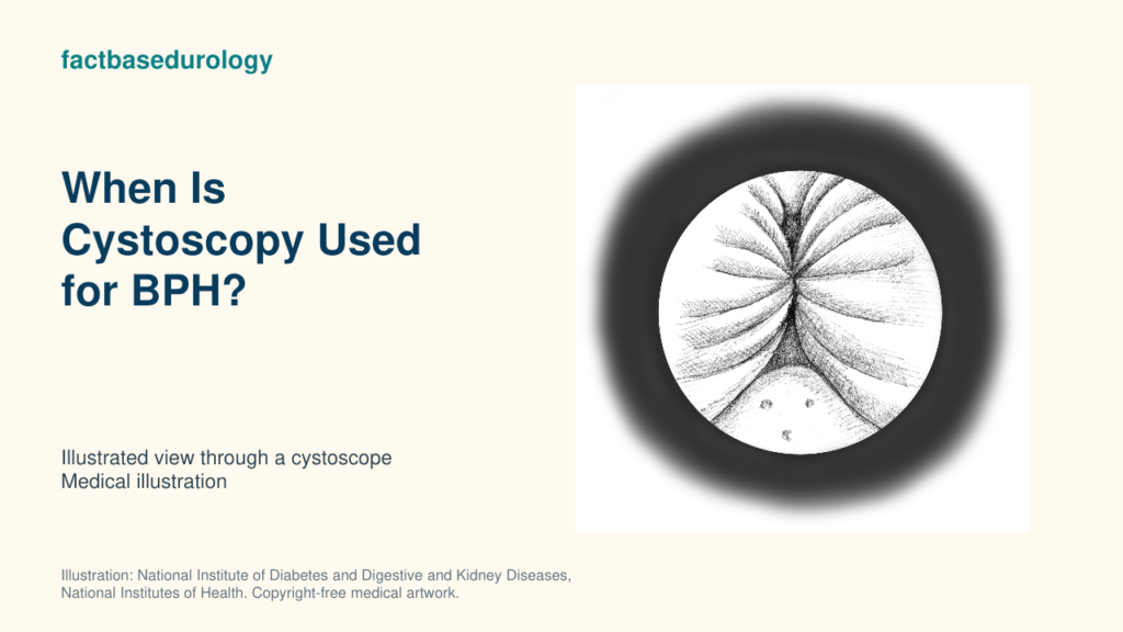
Cystoscopy is not a routine test for every man with suspected benign prostatic hyperplasia (BPH). It is used when directly seeing the urethra, prostate channel, bladder neck or bladder could clarify another diagnosis or change a planned procedure.

01. Cystoscopy: Timing, Indications and Clinical Decision Points
Cystoscopy is considered selectively in BPH evaluation for haematuria, suspected urethral narrowing, bladder-cancer history or anatomy that affects a planned procedure.
Which findings make cystoscopy appropriate to consider?
Cystoscopy is considered for haematuria, stricture risk, bladder lesions or procedure planning because it directly inspects the urethra, prostate outlet and bladder.
| Finding or decision | What cystoscopy can add | Why it matters |
|---|---|---|
| Visible or microscopic haematuria | Direct inspection of urethra and bladder | Looks beyond BPH for bleeding sources. |
| Suspected urethral stricture | Shows the location and calibre of narrowing | A stricture can mimic prostate-related obstruction. |
| History of bladder cancer | Examines bladder lining for recurrence | LUTS should not automatically be attributed to BPH. |
| Recurrent infection, stones or unexplained irritative symptoms | May identify stones, foreign material or mucosal abnormality | Findings can redirect management. |
| Planning selected BPH procedures | Assesses urethra, bladder neck, lateral lobes and median lobe | Anatomy may determine procedural suitability. |
Which symptoms, test results or risk factors affect timing?
Cystoscopy timing depends on haematuria, infection, prior urethral procedures, suspected stricture and cancer history; the underlying clinical concern determines urgency. A poor flow rate or high residual alone usually does not require immediate cystoscopy because neither identifies the mechanism; the test is selected when direct visual anatomy could change the next decision.
02. When Is Cystoscopy Indicated?
Cystoscopy is indicated when direct inspection would clarify a suspected lesion, urethral narrowing or anatomy relevant to BPH treatment.
What prerequisites should be completed before cystoscopy?
Before BPH-related cystoscopy, clinicians review history, medicines, examination and urinalysis; symptom, flow and residual-urine assessment address the broader urinary problem. Symptom scoring, uroflowmetry and post-void residual quantify the functional problem; PSA, renal function or imaging are added when clinically indicated. Active urinary infection should be identified and managed before elective instrumentation.
Which patients may not need cystoscopy immediately?
Men with uncomplicated stable LUTS and no haematuria, stricture risk, bladder-cancer history or procedure-specific anatomical question often do not need immediate cystoscopy. The EAU recommends urethrocystoscopy when findings may change diagnosis or treatment, not simply because BPH is suspected.
03. Cystoscopy: Selection Criteria, Prerequisites and Alternatives
Cystoscopy selection depends on a question that direct inspection can answer; non-invasive tests assess other components of prostate and bladder evaluation.
What alternatives can be considered before cystoscopy?
Alternatives to cystoscopy include urinalysis, ultrasound, uroflowmetry and PVR measurement; each answers a different question and does not replace inspection of a suspected lesion.
Urinalysis evaluates infection and blood; ultrasound measures bladder, residual urine and prostate anatomy; uroflowmetry records the flow pattern; retrograde urethrography can map a suspected stricture; and pressure-flow urodynamics tests functional obstruction. These methods answer different questions and are not interchangeable with direct visual inspection.
What does the indication for cystoscopy not guarantee about outcome?
An indication for cystoscopy does not guarantee that BPH caused symptoms or that an outlet procedure will help; visible anatomy does not establish functional obstruction.
04. What Should Happen Before Proceeding With Cystoscopy?
Before cystoscopy, clinicians confirm its purpose, address infection and medication considerations, and explain preparation, discomfort and procedure risks.
When should the decision for cystoscopy be reassessed?
The cystoscopy decision is reassessed when infection develops, medication management remains unresolved or non-invasive findings have already answered the clinical question. After the procedure, fever, inability to urinate, worsening bleeding or severe pain requires prompt clinical contact; mild short-lived burning and light bleeding can occur.
How does cystoscopy connect to the next treatment or diagnostic step?
Cystoscopy findings guide treatment of strictures, stones or bladder lesions and define anatomy relevant to BPH procedures; functional uncertainty sometimes requires pressure-flow assessment. Anatomy relevant to a BPH procedure—such as a prominent median lobe, bladder-neck configuration or urethral narrowing—can refine procedural selection. A normal examination does not end evaluation when objective flow, residual urine or symptoms remain concerning. The next decision returns to the complete BPH treatment assessment, not the cystoscopic image alone.
Summary
- Cystoscopy is selective, not routine, in uncomplicated BPH evaluation.
- It is useful for haematuria, stricture risk, bladder-cancer history and procedure planning.
- Direct visual anatomy cannot by itself prove functional outlet obstruction.
- The test should be performed only when its result can change diagnosis or management.
Educational disclaimer: This article provides general medical education and cannot diagnose an individual condition or replace assessment by a qualified clinician.
Watch: How BPH is diagnosed
Cystoscopy inspects the urethra and bladder within BPH evaluation. FactBasedUrology explains how symptoms, examination and selected tests contribute to the broader diagnostic process in this video.