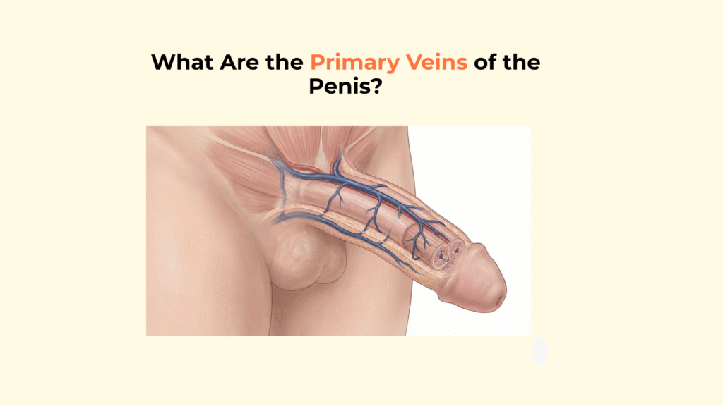
What Are the Primary Veins of the Penis?
The primary veins of the penis are classified into two distinct systems based on their depth relative to Buck’s fascia: the superficial dorsal vein, which drains the skin, and the deep dorsal vein, which drains the erectile tissue.
Distinguishing between these two systems is crucial for understanding both cosmetic appearance (visible blue veins) and functional health (erection maintenance). This guide details the specific anatomy of the penile venous system, mapping the drainage territories, destinations, and functional roles of the superficial and deep veins.
Important Medical Disclaimer
This information is for educational purposes only and is not a substitute for professional medical advice, diagnosis, or treatment. Consult with a qualified healthcare provider regarding any medical condition or concerns about your health.
Key Vascular Facts: Primary Veins
- The Systems: Penile veins are divided into a Superficial System and a Deep System.
- The Boundary: The dividing line is Buck’s Fascia. Superficial is above it; Deep is below it.
- Superficial Dorsal Vein: Drains the skin and foreskin. Empties into the leg veins (Saphenous).
- Deep Dorsal Vein: Drains the glans and corpora cavernosa. Empties into the pelvic plexus.
- Functional Role: Only the Deep System is involved in maintaining an erection via the veno-occlusive mechanism.
How Are the Primary Veins of the Penis Classified Anatomically?
The primary veins of the penis are classified anatomically based on their relationship to the deep fascia (Buck’s fascia), dividing them into the superficial and deep venous systems.
The Superficial Venous System
The Superficial Venous System consists mainly of the Superficial Dorsal Vein, which lies within the loose areolar tissue between the Dartos fascia and Buck’s fascia. It drains the skin, prepuce (foreskin), and subcutaneous tissue. This system has minimal influence on erection quality; these are often the prominent blue veins visible under the skin.
The Deep (Intermediate) Venous System
The Deep Venous System is centered on the Deep Dorsal Vein, which lies deep to Buck’s Fascia, running in the dorsal groove between the two corpora cavernosa. It drains the Glans Penis, Corpus Spongiosum, and the distal two-thirds of the Corpora Cavernosa via circumflex veins (curved veins that wrap around the shaft to feed into the deep dorsal vein).
The Deep (Proximal) Venous System
The Deep (Proximal) Venous System comprises the Cavernosal Veins and Crural Veins, which exit directly from the crura (roots) of the penis. They exit directly from the base of the erectile bodies behind the pubic bone and drain the proximal one-third of the erectile tissue.
What Are the Anatomical Destinations of the Primary Veins of the Penis?
The anatomical destinations of the primary veins differ significantly, with the superficial system draining externally toward the leg and the deep system draining internally into the pelvic plexus.
The External Drainage Route (Superficial)
The Superficial Dorsal Vein drains into the External Pudendal Vein, which then empties into the Great Saphenous Vein, part of the leg’s venous system. This anatomical connection explains why penile skin infections can sometimes cause lymphadenopathy (swollen lymph nodes) in the superficial inguinal nodes of the groin.
The Internal Drainage Route (Deep)
The Deep Dorsal Vein passes beneath the pubic arch to drain directly into the Periprostatic Plexus (also known as Santorini’s Plexus). This connection creates a direct route to the pelvic venous system, linking penile drainage directly to pelvic floor health. For a detailed overview of pelvic anatomy, refer to StatPearls on Pelvic Anatomy.
How Do Valves Function Within the Primary Veins of the Penis?
Valves within the primary veins of the penis function to ensure unidirectional blood flow toward the heart, preventing backflow (reflux) that could compromise circulation.
The “Valve-Competence” Role
The Deep Dorsal Vein typically contains valves to direct flow proximally away from the glans. Incompetent valves in the Deep Dorsal Vein can contribute to venous leak (retrograde flow), a cause of erectile dysfunction.
The “Valve-Less” Emissary Veins
In contrast, the tiny emissary veins piercing the tunica albuginea are essentially valveless. Because they lack valves, they rely entirely on the compression of the Tunica Albuginea (veno-occlusive mechanism) to stop outflow during an erection. This explains why structural integrity (the Tunica) matters more than valves for erection maintenance.
Comparative Matrix: Superficial vs. Deep Primary Veins
This table provides a direct comparison of the superficial and deep venous systems of the penis, detailing their location, drainage source, and destination.
| Vein | Fascial Layer | Drains… | Empties Into… |
|---|---|---|---|
| Superficial Dorsal Vein | Above Buck’s Fascia | Skin/Dartos | Ext. Pudendal / Saphenous |
| Deep Dorsal Vein | Below Buck’s Fascia | Glans / Distal Cavernosa | Periprostatic Plexus |
| Cavernosal Veins | Intracrural (Deep Root) | Proximal Cavernosa | Int. Pudendal |
Integrated Vascular Physiology
The deep dorsal vein does not function in isolation, because the blood it drains originates inside the corpora cavernosa , where intracavernosal pressure must rise high enough to mechanically restrict venous outflow during erection.
This venous compression depends on the tensile stiffness of the tunica albuginea , which acts as the non-yielding outer shell that flattens the emissary veins as the erectile bodies expand.
Before venous occlusion can occur, arterial inflow must first expand the sinusoids through the action of the helicine arteries , which regulate resistance at the microvascular entry point of the cavernous tissue.
The deep venous system ultimately connects to the pelvic circulation through the same structural foundation that anchors the root of the penis, as described in the pelvic attachment anatomy , where the proximal venous channels pass beneath the pubic arch.
Sensory feedback from the dorsal aspect of the penis travels alongside the deep venous structures via the dorsal penile nerve , which explains why surgical trauma in this compartment can affect both sensation and venous drainage.
If venous outflow fails to restrict properly, blood escapes faster than it enters, producing the classic pressure-loss pattern seen in erectile dysfunction , despite otherwise intact arterial inflow.
Structural diseases that deform the tunica, such as Peyronie’s disease , can distort the normal geometry of the deep dorsal and emissary veins, leading to asymmetric venous occlusion.
Proximally, blood drained by the cavernosal and crural veins transitions into the broader penile vascular pathway , where penile venous return integrates into the pelvic venous plexus and systemic circulation.
The mechanical regulation of venous outflow is impossible without simultaneous relaxation of the surrounding erectile smooth muscle tissue , which allows the cavernous spaces to expand and generate the compressive force required for veno-occlusion.
Conclusion
In conclusion, the primary veins of the penis form a dual drainage system separated by Buck’s fascia, with the superficial system handling skin drainage and the deep system managing the critical blood flow dynamics of erection.
At Factbasedurology, we believe that detailed anatomical knowledge is the foundation for understanding your body’s function. This complex vascular network ensures both the health of the tissues and the mechanics of sexual function.