What Is the Vascular Pathway for Blood Flow into the Penis?
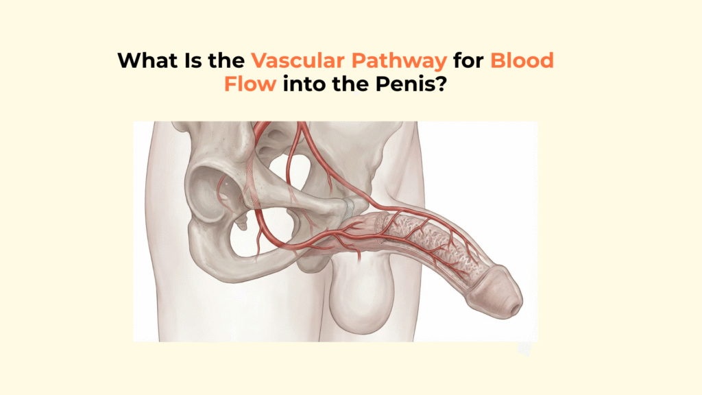
The vascular pathway for blood flow into the penis begins at the abdominal aorta, travels through the internal iliac and internal pudendal arteries, and terminates by branching into the cavernosal, dorsal, and bulbourethral arteries.
This complex route, involving multiple junctions and a critical passage through the pelvis, ensures the high-pressure delivery required for erection. If insufficient blood volume reaches the tissue, the pressure gradient collapses, leading to erectile dysfunction. This guide provides a step-by-step anatomical map of the penile vascular pathway, tracing the blood from the heart to the microscopic sinusoids.
This information is for educational purposes only and is not a substitute for professional medical advice, diagnosis, or treatment. Consult with a qualified healthcare provider regarding any medical condition or concerns about your health.
Key Pathway Facts: Penile Blood Flow
- ● The Source: The pathway starts at the Abdominal Aorta and splits into the Common Iliac Arteries.
- ● The Pelvic Route: The Internal Iliac Artery feeds the Internal Pudendal Artery, the main vessel for the perineum.
- ● The Critical Tunnel: The artery passes through Alcock’s Canal, a common site for compression injury.
- ● The Junction: The Common Penile Artery trifurcates (splits) into three terminal branches.
- ● The Destination: The Cavernosal Artery feeds the Helicine Arteries, which dump blood into the sinusoids.
What Is the Systemic Origin of the Vascular Pathway to the Penis?
The systemic origin of the vascular pathway to the penis is the abdominal aorta, which descends through the abdomen before branching into the specialized pelvic arteries.
From Aorta to Pelvis (The Macro-Pathway)
The macro-pathway involves a sequential bifurcation: the Abdominal Aorta splits into the Common Iliac Arteries, which then divide into the Internal Iliac Artery (Hypogastric Artery).
- Step 1: Abdominal Aorta bifurcates at the L4 vertebra.
- Step 2: Common Iliac splits at the pelvic brim, a key landmark in pelvic attachment anatomy.
- Step 3: The Anterior Division of the Internal Iliac gives rise to the Internal Pudendal Artery.
The Critical “Alcock’s Canal” Transit
The Internal Pudendal Artery takes a complex course, exiting the pelvis via the greater sciatic foramen and re-entering via the lesser sciatic foramen to travel through Alcock’s Canal (pudendal canal). It travels along the lateral wall of the ischiorectal fossa.
This is a common site for compression, known as Pudendal Nerve Entrapment or ‘cyclist syndrome,’ which can affect the vascular pathway and sensation. (Source: StatPearls).
How Does the Vascular Pathway Terminate and Branch into the Penis?
Just below the urogenital diaphragm, the Internal Pudendal Artery terminates by becoming the Common Penile Artery, which then undergoes a “trifurcation event” to supply the specific penile structures.
The “Common Penile Artery” Junction
The Common Penile Artery splits into three distinct pathways: the Bulbourethral Artery (supplying the bulb), the Dorsal Artery (supplying the glans), and the Cavernosal Artery (supplying the erectile tissue).
- Bulbourethral Artery: Supplies the Bulb and Corpus Spongiosum.
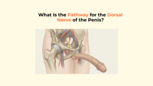
- Dorsal Artery: Supplies the skin, fascia, and Glans, coursing alongside the dorsal penile nerve.
- Cavernosal Artery: Supplies the Corpora Cavernosa.
The “Cavernosal” Inflow Route (The Erection Path)
The Cavernosal Artery enters the Crura (roots) of the penis and runs longitudinally through the center of the corpora cavernosa (erectile tissue) to drive erection.
Mechanism Logic: The central positioning of the Cavernosal Artery allows for uniform distribution of blood, ensuring the entire cylinder expands simultaneously.
What Is the Micro-Vascular Pathway from Artery to Erectile Tissue?
The micro-vascular pathway involves the branching of the Cavernosal Artery into numerous Helicine Arteries, which open directly into the vascular spaces.
The Helicine Artery Branching
The Cavernosal Artery gives off numerous helicine arteries, which act as the final resistance vessels before blood enters the Lacunar Spaces (Sinusoids). Imagine a main highway (Cavernosal) with hundreds of exit ramps (Helicine) leading into parking lots (Sinusoids). These vessels dilate during arousal, a process dependent on the relaxation of erectile smooth muscle tissue. For blood to remain trapped inside, venous outflow is restricted via the penile venous system.
The “Nutrient Pathway” Exception
A distinct set of short “nutrient arteries” branches off to supply the collagenous framework (trabeculae) and the tunica albuginea directly, ensuring tissue health independent of erection. Structural disorders here, such as Peyronie’s disease, can deform this geometry. Not all blood goes to the sinusoids; this parallel pathway maintains tissue viability.
Visualizing the Complete Vascular Pathway Flowchart
This step-by-step flowchart traces the complete journey of a blood cell from the heart to the erectile tissue.
- Abdominal Aorta
- Common Iliac Artery
- Internal Iliac Artery (Anterior Division)
- Internal Pudendal Artery (via Alcock’s Canal)
- Common Penile Artery
-
Trifurcation:
- 6a. Bulbourethral A. (to Spongiosum)
- 6b. Dorsal A. (to Glans)
- 6c. Cavernosal A. (Deep Artery)
- Helicine Arteries
- Lacunar Spaces (Sinusoids)
[Checklist] Tracing the Vascular Pathway to the Penis
Use this checklist to verify your understanding of the penile vascular route.
- Systemic Origin: Is the Internal Iliac (Anterior Division) identified as the source?
- Transit Route: Is Alcock’s Canal noted as the passage for the Pudendal Artery?
- The Split: Is the Common Penile Artery identified as the branching point?
- Erection Route: Is the Cavernosal Artery -> Helicine Artery path identified as the specific route for rigidity?
- Target: Do the vessels ultimately terminate in the Lacunar Spaces (sinusoids)?
Glossary of Anatomical Terms
To ensure full clarity, this glossary defines the key anatomical terms used throughout this guide on the vascular pathway.
| Term | Definition |
|---|---|
| Abdominal Aorta | The largest artery in the body, which supplies oxygenated blood to the abdominal and pelvic organs. |
| Internal Iliac Artery | The main artery of the pelvis that supplies blood to the reproductive organs, buttocks, and pelvic muscles. |
| Internal Pudendal Artery | A branch of the internal iliac artery that supplies blood to the external genitalia and perineum. |
| Alcock’s Canal | An anatomical tunnel in the pelvis through which the internal pudendal artery and nerve pass. |
| Common Penile Artery | The terminal branch of the internal pudendal artery before it splits into the three main penile arteries. |
| Sinusoids | The vascular spaces within the erectile tissue that fill with blood during an erection. |
Conclusion
In conclusion, the vascular pathway to the penis is a highly specific and regulated route, ensuring that blood is delivered precisely where it is needed—whether for tissue nutrition via the dorsal artery or for hydraulic rigidity via the cavernosal artery. The journey from the Aorta through Alcock’s Canal to the Common Penile Artery trifurcation represents a critical anatomical sequence where blockages or compression can significantly impact function.
At Factbasedurology, we believe that detailed anatomical knowledge empowers you to better understand your body’s function. Tracing this pathway reveals the intricate plumbing that makes sexual function possible.
3 Responses