
Prostate anatomy · BPH growth · Updated August 26, 2026
What Is the Prostate Transition Zone? Anatomy, BPH Growth and Clinical Significance
The transition zone is a small inner region of the prostate with outsized clinical importance. Its position around the urethra explains why tissue growth there can alter urine flow, while its mixed glandular and stromal composition explains why benign nodules can be difficult to distinguish from cancer on imaging.
Where is the transition zone located?
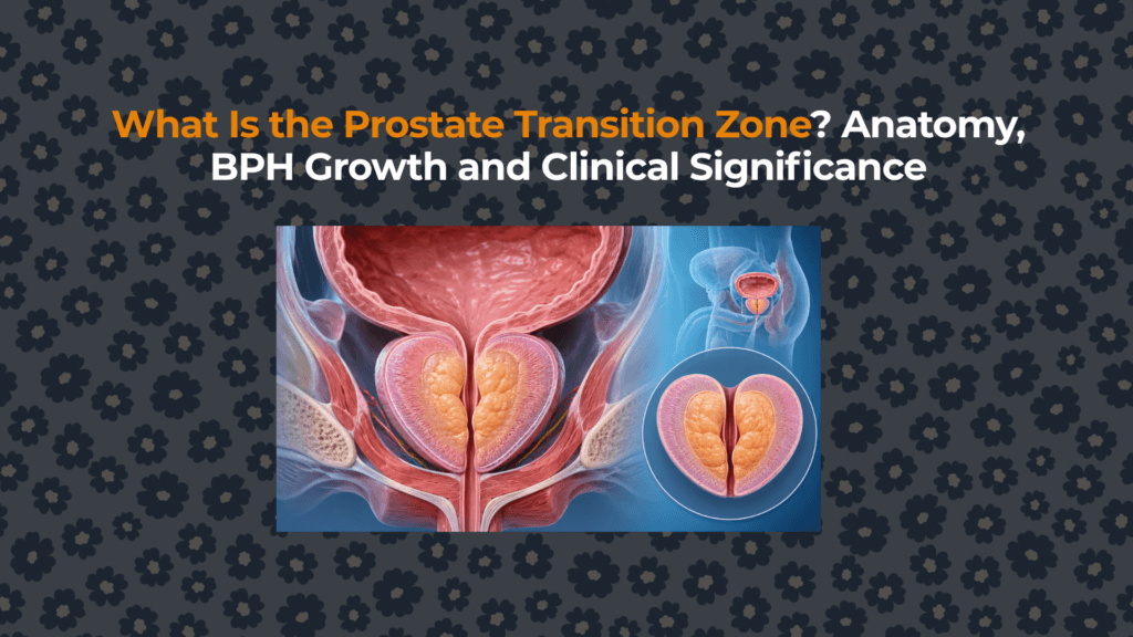
The transition zone forms an anterolateral, horseshoe-like ring around the upper prostatic urethra. It lies inside the peripheral zone and below the bladder neck. Inferiorly, its relationship is commonly described relative to the verumontanum—the ridge where the ejaculatory ducts enter the prostatic urethra.
This location is easier to understand after reviewing the complete map of the peripheral, central and transition zones. The transition zone is not the entire “inner gland,” and it is not identical to the median lobe. Those terms describe overlapping but different anatomical concepts.
How large is the transition zone normally?
In the classic McNeal model, the transition zone accounts for approximately 5% of prostate glandular tissue in a young adult—often only a little over 1 mL in a prostate near 20–25 mL. This is an anatomical teaching estimate, not a universal reference interval.
Age, hormones, stromal-epithelial signaling and individual biology can change the zone substantially. Once BPH develops, transition-zone volume may account for a much larger fraction of total prostate volume. Therefore, a radiology report describing an enlarged transition zone should be interpreted using the person’s total volume, morphology, symptoms, urine-flow findings and other clinical information.
Why does BPH develop in the transition zone?
BPH is a histological process involving noncancerous proliferation of epithelial and stromal cells. It arises mainly in transition-zone and periurethral tissue rather than uniformly throughout the prostate. Nodules may be predominantly glandular, predominantly stromal, or mixed.
The exact initiating mechanism is multifactorial. Aging and continued androgen signaling—particularly local conversion of testosterone to dihydrotestosterone—are important, but BPH is not simply “too much testosterone.” Local growth factors, inflammatory signaling, extracellular matrix and epithelial–stromal interactions contribute to tissue remodeling.
The dedicated BPH hub will distinguish histologic growth from anatomical enlargement and functional obstruction: BPH and enlarged-prostate evidence.
How can transition-zone growth affect urination?
Urine must pass from the bladder through the prostatic urethra. Expanding tissue around this channel can increase outlet resistance. Possible consequences include weak stream, hesitancy, intermittency, incomplete emptying and urinary retention.
Symptoms are not a direct volume gauge. A relatively small median-lobe or intravesical protrusion may cause substantial obstruction, while a larger gland may produce limited symptoms. Bladder muscle strength, urethral resistance, autonomic tone, medications, neurologic disease and fluid patterns also influence lower urinary tract symptoms.
How is transition-zone volume measured?
Ultrasound or MRI can estimate total prostate and transition-zone dimensions. A common approximation treats the region as an ellipsoid: width × height × length × about 0.52. The transition-zone index is calculated as transition-zone volume divided by total prostate volume.
These measurements are estimates affected by imaging plane, boundary selection and irregular BPH nodules. Research has evaluated whether transition-zone index correlates with symptoms, maximum flow and bladder pressure, but no single ratio replaces clinical assessment. The forthcoming prostate-volume measurement guide explains ellipsoid assumptions and measurement error.
| Measure | Calculation or observation | What it can contribute | What it cannot prove |
|---|---|---|---|
| Total prostate volume | Three dimensions × ≈0.52 | Overall gland size; treatment planning | Cause or severity of symptoms |
| Transition-zone volume | Inner-zone dimensions × ≈0.52 | Extent of inner-gland enlargement | Definite obstruction or cancer |
| Transition-zone index | TZ volume ÷ total volume | Proportion occupied by transition zone | A standalone treatment decision |
| Intravesical protrusion | Protrusion into bladder measured on imaging | Outlet geometry and median-lobe relevance | Bladder contractility |
| Post-void residual | Urine remaining after voiding | Emptying efficiency at that time | That BPH is the sole cause |
What does the transition zone look like on MRI?
A normal young transition zone is relatively compact. With BPH it becomes enlarged and heterogeneous because glandular nodules may appear relatively T2 bright while smooth-muscle-rich stromal nodules can be T2 dark. Encapsulated, circumscribed nodules often support a benign interpretation, but appearance varies.
Transition-zone cancer is less common than peripheral-zone cancer but clinically important. It may be more anterior and less accessible to digital rectal examination. PI-RADS evaluates transition-zone lesions primarily with T2-weighted morphology, with diffusion findings contributing to assessment. Stromal BPH can mimic cancer through low T2 signal, diffusion restriction and enhancement, so MRI does not substitute for integrated risk assessment or histology when biopsy is indicated.
Transition zone vs peripheral zone
| Attribute | Transition zone | Peripheral zone |
|---|---|---|
| Position | Around proximal urethra | Posterior and lateral outer gland |
| Young-adult proportion | ≈5% glandular tissue | ≈70% glandular tissue |
| Dominant benign process | BPH | Inflammation and other benign changes |
| Share of cancers | Often cited around 20–25% | Often cited around 60–75% |
| MRI challenge | Heterogeneous BPH nodules can mimic cancer | Inflammation/hemorrhage can mimic cancer |
| DRE access | Often less directly palpable | Posterior portion more accessible |
For the complementary outer-gland perspective, see the peripheral-zone anatomy and cancer-relevance guide.
Summary
The transition zone is a small paired region wrapped around the proximal prostatic urethra. It starts near 5% of glandular tissue in many young adults but can enlarge dramatically because it is the main site of BPH. Its growth can alter outlet geometry, yet size alone does not diagnose obstruction or explain every urinary symptom. MRI can characterize transition-zone structure, but benign stromal and glandular nodules may overlap with cancer appearances. Accurate interpretation requires symptoms, examination, PSA context, urine-flow or residual testing, imaging and sometimes biopsy.
This article is educational and does not diagnose urinary symptoms or interpret an individual MRI.


