
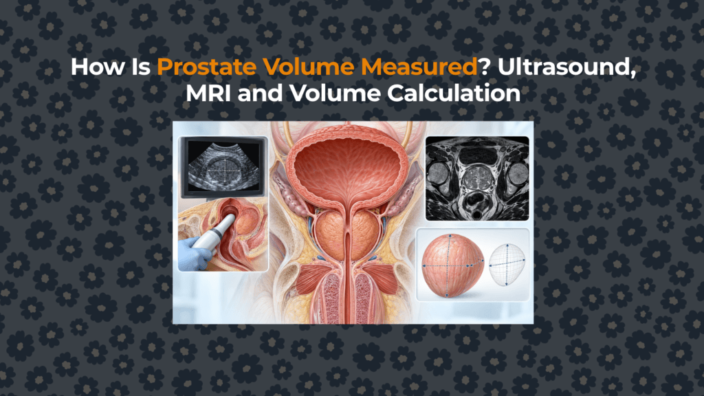
How Is Prostate Volume Measured? Ultrasound, MRI and Volume Calculation
Prostate volume is not read directly from a scan. It is estimated from a boundary drawn around a three-dimensional gland.
01. What exactly is a prostate-volume result?
A report in millilitres or cubic centimetres is a geometric estimate of the tissue enclosed by an interpreted prostate boundary. It is not the same as pathology weight, transition-zone volume or obstructing tissue volume. One millilitre equals one cubic centimetre, so reports using mL and cm³ are numerically comparable.
Start with the prostate anatomy map if the base, apex, capsule or zones are unfamiliar. For interpretation of the resulting number—not its acquisition—use the separate normal prostate-size reference guide.
02. How does the ellipsoid formula turn three diameters into volume?
The coefficient approximates π/6, converting the surrounding rectangular box into a prolate ellipsoid. A gland measured as 5.0 × 4.0 × 4.0 cm therefore gives 41.9 mL. Because all three axes are multiplied, a small error in more than one diameter compounds rather than cancels.
03. Why can the formula fail in an irregular or median-lobe gland?
The ellipsoid model assumes a smooth, symmetric solid. BPH nodules, asymmetric transition-zone growth and intravesical protrusion violate that assumption. Axis placement also matters: an oblique rather than true midline length can enlarge the product.
What does comparison with planimetry show?
In a retrospective brachytherapy cohort of 138 glands, only 13.3% of conventional ellipsoid estimates were within 10% of planimetry; the formula underestimated volume by 17% on average (95% CI 14%–19%). A bullet-shape coefficient, π/4.8, placed 75% within 10%. This does not make the bullet formula universally superior: the cohort was selected for brachytherapy and performance improved particularly below 55 cm³.
04. How does transabdominal ultrasound measure the gland?
Transabdominal ultrasound (TAUS) images the prostate through the lower abdomen, usually with a filled bladder as an acoustic window. It is non-invasive and readily available, but depth, body habitus, bladder filling and limited definition of the apex or base can alter boundary selection.
05. How is transrectal ultrasound different?
TRUS places the probe near the prostate through the rectum, improving spatial access and supporting biopsy or procedural planning. It still depends on probe angle, pressure and chosen axes. In an EAU-cited series of 640 men, TRUS volumes averaged 8% smaller than MRI; that group result is not a universal conversion factor.
The probe’s relationship to the gland is mapped in the prostate-location article.
06. What does MRI add?
MRI provides multiplanar soft-tissue contrast and supports either the same ellipsoid calculation or slice-by-slice planimetry/segmentation. In one reproducibility study summarized by EAU, seven radiologists measuring 40 MRI scans achieved intra- and inter-reader intraclass correlations above 0.90 across two ellipsoid methods and manual planimetry. High reproducibility means readers rank and repeat measurements consistently; it does not prove zero absolute error.
Which MRI landmarks define the length?
The craniocaudal axis should be established on a true midsagittal T2 image from the bladder–prostate junction to the apical margin. Width is measured on the axial plane at the gland’s largest transverse diameter; the anteroposterior axis should be taken in the same plane. A median lobe projecting into the bladder can make the base ambiguous, while indistinct apical tissue can shorten or lengthen the chosen boundary. Reporting the method is therefore part of the measurement—not optional metadata.
When is segmentation preferable?
Planimetry traces the gland on successive slices and sums area × slice thickness. It preserves irregular contour better than one ellipsoid, but requires more time, software and boundary decisions. Semiautomated segmentation reduces manual work but still needs quality control at the base, apex and around protruding nodules.
07. Which method is most accurate against a specimen reference?
| Evidence unit | TAUS | MRI | Boundary |
|---|---|---|---|
| Guo et al., 106 prostatectomy patients | r=0.838; ICC=0.83 | r=0.914; ICC=0.90 | Selected cancer cohort; ex-vivo specimen is not identical to living tissue |
| Estimate within ±20% of specimen | 65/106 (61%) | 87/106 (82%) | Shows agreement distribution, not guaranteed error for one patient |
| Mean direction | +2.4 mL | −1.7 mL | Averages can conceal wide individual differences |
08. Why can a volume error change PSA density?
PSA density equals serum PSA divided by prostate volume. With PSA fixed at 6 ng/mL, a 30 mL estimate gives 0.20 ng/mL/cc, while 40 mL gives 0.15. Thus a boundary or axis difference propagates directly into the denominator and can move a result across a decision threshold. PSA density must be interpreted within the MRI and clinical-risk pathway, not as an isolated calculation.
09. How should two different volume reports be compared?
| Check | Why it matters |
|---|---|
| Same modality and route? | TAUS, TRUS and MRI use different viewing geometry. |
| Same three planes? | Oblique or non-midline axes change the product. |
| Same method? | Ellipsoid and segmentation do not have identical assumptions. |
| Same gland boundary? | Base, apex and median-lobe inclusion may differ. |
| Large enough change? | Small differences can reflect measurement variability rather than growth. |
When should volume be repeated or confirmed?
Confirmation is most useful when the number would change PSA-density interpretation, medicine eligibility or procedure selection; when the gland is irregular; or when two reports disagree beyond an expected method difference. Prefer the same modality, acquisition route, formula and landmark convention for serial comparison. A repeat solely to chase a small numerical difference may add false precision.
When the question is ageing rather than technique, use the longitudinal prostate-change evidence. For symptoms, volume must remain separate from the size–symptom relationship and the BPH evidence hub.
Clinical interpretation boundary
Evidence sources
- EAU prostate-cancer diagnostic evaluation: volume estimation and PSA density.
- Guo et al. 2023: TAUS and MRI versus prostatectomy specimen volume.
- Wasserman et al.: MRI boundary landmarks and measurement guide.
- MacMahon et al.: ellipsoid versus bullet-shape formula.
- Systematic review of imaging accuracy across 7,768 patients.
- Review of ellipsoid, planimetry and segmentation methods.
- Ultrasound and MRI agreement study.


