What Is the Primary Tissue That Makes Up the Corpora Cavernosa?
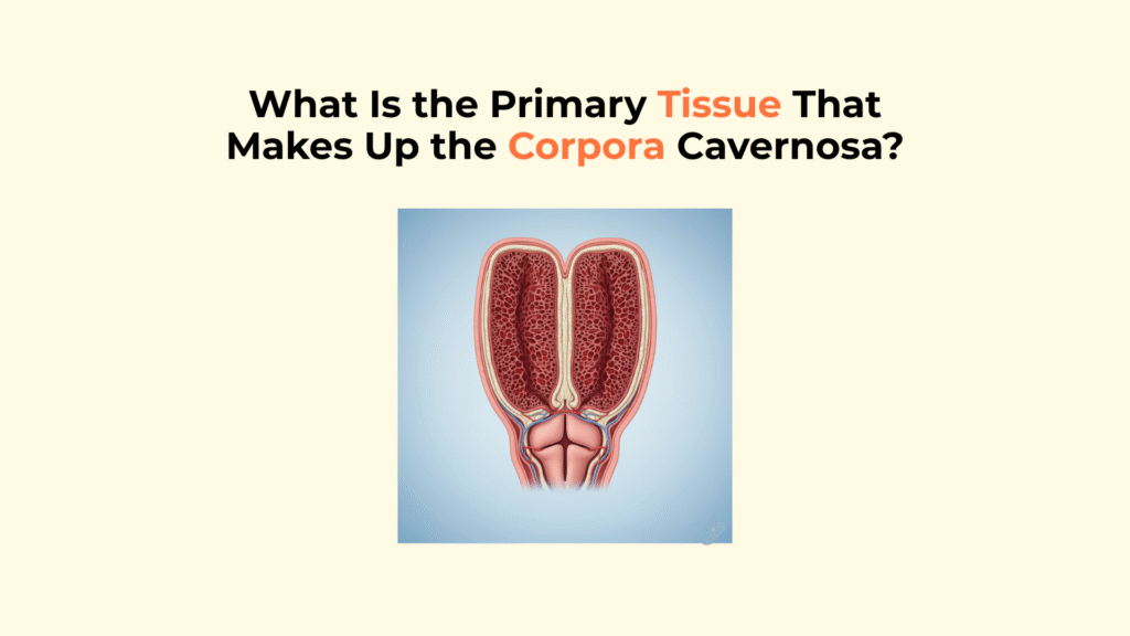
The primary tissue making up the Corpora Cavernosa is a specialized form of erectile tissue, a unique sponge-like network composed of smooth muscle fibers, fibrous connective tissue (trabeculae), and interconnected vascular spaces called sinusoids.
This unique erectile tissue structure is fundamentally different from skeletal muscle; it is designed to function like a complex hydraulic sponge, rapidly filling with blood to produce a rigid erection.
This guide provides a detailed, medically accurate explanation of the ‘corpora cavernosa tissue,’ its unique structure, and the precise physiological mechanism it uses to create an erection. Understanding the specific components of this tissue, from the trabeculae smooth muscle to the sinusoids, is essential for grasping how an erection occurs and how certain medical conditions can affect it.
Important Medical Disclaimer
This information is for educational purposes only and is not a substitute for professional medical advice, diagnosis, or treatment. Consult with a qualified healthcare provider regarding any medical condition or concerns about your health.
Key Takeaways on Corpora Cavernosa Tissue
- What it is: The Corpora Cavernosa are the two main erectile chambers of the penis, composed of a sponge-like erectile tissue.
- Key Components: This tissue is a network of sinusoids (vascular spaces) supported by a framework of trabeculae (which contain smooth muscle).
- How Erections Work: An erection occurs when smooth muscle in the trabeculae relaxes, allowing blood to flood and fill the sinusoids.
- The Outer Sheath: This tissue is encased by the Tunica Albuginea, a tough sheath that traps blood at high pressure to create rigidity.
What Is the Structure of the Corpora Cavernosa’s Erectile Tissue?
Structurally, the erectile tissue within the Corpora Cavernosa consists of two main components: a three-dimensional framework of trabeculae and the vascular spaces known as sinusoids situated between them. This internal architecture is what gives the tissue corpora cavernosa its unique erectile properties.
What Are the Trabeculae?
The trabeculae are an intricate, three-dimensional network of interconnected beams and columns that form the structural framework of the Corpora Cavernosa’s erectile tissue. They are composed primarily of smooth muscle fibers interwoven with fibrous connective tissue, including collagen for strength and elastin for flexibility. The smooth muscle component within the trabeculae is the “gatekeeper” that controls blood flow into the sinusoids.
What Are the Sinusoids (or Lacunar Spaces)?
The sinusoids, also known as lacunar spaces, are the irregular, interconnected vascular cavities located between the trabeculae within the Corpora Cavernosa. These are essentially expandable, sponge-like blood spaces. Lined by a thin layer of specialized cells called the endothelium, these sinusoids are the spaces that expand dramatically and fill with arterial blood to produce an erection.
How Does the Corpora Cavernosa’s Tissue Function During an Erection?
The Corpora Cavernosa’s tissue functions during an erection via the neurologically controlled relaxation of smooth muscle within the trabeculae, which allows the sinusoids to rapidly engorge with blood. This process is a key part of penile hemodynamics.
How Do the Trabeculae Control Blood Flow?
The trabeculae control blood flow into the sinusoids through the contraction or relaxation of their integrated smooth muscle fibers.
- Flaccid State: In the non-erect state, the smooth muscle within the trabeculae is tonically contracted (due to sympathetic nerve signals). This contraction restricts arterial inflow and keeps the sinusoids relatively empty.
- Erect State: During arousal, parasympathetic nerve signals trigger the release of Nitric Oxide (NO), which causes this smooth muscle to relax.
How Does This Tissue Create Rigidity?
This erectile tissue creates rigidity when smooth muscle relaxation allows arterial blood to rapidly fill and expand the sinusoids, trapping the blood under high pressure and stiffening the entire structure.
The physiological sequence is precise: Nitric Oxide (NO) signals the trabeculae smooth muscle to relax, allowing the sinusoids to fill with blood.
This expansion compresses the draining veins against the outer sheath, trapping the blood and causing a rigid erection.
This hemodynamic event increases blood flow from a flaccid rate of 2-6 mL/min to an erect rate of 25-60 mL/min (Source: NIH). This is a hydraulic event, fundamentally dependent on smooth muscle relaxation, not skeletal muscle contraction.
What Role Does the Tunica Albuginea Play with the Corpora Cavernosa’s Tissue?
The Tunica Albuginea plays a critical synergistic role with the Corpora Cavernosa’s internal erectile tissue by acting as a strong, relatively inelastic outer sheath that contains the expanding tissue and generates high pressure.
What Is the Tunica Albuginea?
The Tunica Albuginea is the thick, tough, bi-layered fibrous sheath composed primarily of collagen that tightly encases each of the two Corpora Cavernosa. Its bi-layered structure (inner circular and outer longitudinal fibers) contributes to its immense strength and ability to withstand high pressure.
How Does It Create Hardness (The Veno-Occlusive Mechanism)?
The Tunica Albuginea creates the hardness of an erection by resisting the expansion of the blood-filled Corpora Cavernosa tissue, which activates the veno-occlusive mechanism.
The “veno-occlusive mechanism” is the process where the expanding sinusoids compress the draining veins (subtunical venules) against the rigid Tunica Albuginea.
This trapping of blood raises the intracavernosal pressure to approximately 100 mmHg, which is what confers the rigidity and hardness of a functional erection.
This is the specific tissue that develops inelastic scar tissue (plaque) in Peyronie’s disease, which illustrates the tunica albuginea’s role for a straight erection (Source: Mayo Clinic).
Summary Matrix: Key Tissues of the Corpora Cavernosa
This table provides a concise summary comparing the composition and functional state of the key tissue components within and surrounding the Corpora Cavernosa during erection.
| Tissue/Structure | Composition | Primary Function | State During Erection |
|---|---|---|---|
| Trabeculae | Smooth muscle & fibrous tissue | Forms structural framework; controls blood flow | Relaxed |
| Sinusoids | Endothelium-lined spaces | Fill with blood | Engorged |
| Tunica Albuginea | Dense fibrous tissue (collagen) | Traps blood under high pressure | Stretched and taut |
For a full overview of penile anatomy, including how the erectile tissue interfaces with visible structures, see our comprehensive guide to penile anatomy.
While exploring the internal framework, it helps to also review the detailed anatomy of the visible parts of the penis, so you can understand how internal and external structures relate.
The erectile process heavily involves structures like the corpora cavernosa, which are central to how rigidity is achieved.
For readers asking whether the penis is a muscle, our article on why the penis is not a muscle clarifies the distinction between skeletal muscle and erectile tissue.
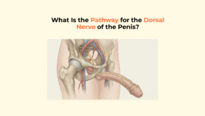
If you’d like to dive into neurovascular control of penile function, explore our section on the dorsal nerve of the penis and how it supports sensation.
Conclusion: A Specialized Hydraulic Tissue
In conclusion, the Corpora Cavernosa are composed not of muscle in the conventional sense, but of a highly specialized erectile tissue designed for a hydraulic function.
This tissue’s composition is an intricate framework of smooth muscle and fibrous tissue (trabeculae) that defines expandable vascular spaces (sinusoids). This internal spongy tissue works in direct conjunction with the tough outer Tunica Albuginea. Together, they form a sophisticated hydraulic system where smooth muscle relaxation allows rapid blood engorgement within a contained space, generating the high pressure and rigidity of an erection. This elegant hydraulic system, dependent on smooth muscle relaxation and healthy blood flow, is a marvel of physiological engineering.
At Factbasedurology, we believe that understanding this complex anatomy is the first step to appreciating and protecting your vascular health.
Glossary of Clinical Terms
To ensure full clarity, this glossary defines the key anatomical terms used throughout this guide on the corpora cavernosa tissue.
| Term | Definition |
|---|---|
| Corpora Cavernosa | The two main, parallel columns of erectile tissue in the penis shaft that hold 90% of the blood and create a rigid erection. |
| Erectile Tissue | A type of spongy tissue rich in vascular spaces (sinusoids) that fills with blood to cause an erection. |
| Sinusoids (Lacunar Spaces) | The expandable, interconnected vascular spaces within the erectile tissue that fill with blood. |
| Trabeculae | The structural framework of smooth muscle and connective tissue that separates the sinusoids. |
| Tunica Albuginea | The strong, flexible, bi-layered sheath that traps blood at high pressure (over 100 mmHg) to maintain a rigid erection. |
| Smooth Muscle | Involuntary muscle found in blood vessels and the trabeculae. Its relaxation is the key event that causes an erection. |
| Nitric Oxide (NO) | The primary signaling molecule that triggers the relaxation of smooth muscle, initiating the erection process. |
13 Responses