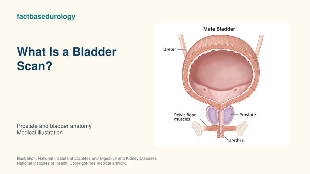
A bladder scan is a quick, non-invasive ultrasound measurement of urine volume in the bladder. In men with lower urinary tract symptoms, it is most often performed immediately after urination to estimate the post-void residual (PVR)—the urine left behind.

01. Bladder Scan: Measurement, Calculation and Interpretation
A bladder scan estimates urine volume non-invasively with ultrasound; a scan immediately after urination measures post-void residual urine.
How is a bladder scan measured or calculated?
A bladder scan places an ultrasound probe over the lower abdomen, usually with gel, to estimate bladder volume; post-void scanning follows urination promptly. While the patient usually lies on the back, gel is placed above the pubic bone and the scanner is aimed toward the bladder. The device locates the fluid-filled bladder, samples it in more than one plane and applies a manufacturer-specific algorithm to estimate volume.
The scanner does not drain the bladder or directly collect urine. Its displayed result is a calculated estimate based on the detected bladder outline. Repositioning and repeating the scan can help when the bladder is not centred or the reading is unexpected.
Which units, thresholds or scoring rules are used for a bladder scan?
A bladder scan reports estimated volume in millilitres; its residual-urine result has no universal BPH treatment threshold. There is no “bladder-scan score.” Any clinical threshold refers to the PVR result, not to the scanner itself, and must be interpreted with symptoms, voided volume, timing and other findings.
| Finding | What the evidence supports | What it does not establish |
|---|---|---|
| PVR ≥50 mL | In EAU-cited evidence, this threshold had a 63% positive predictive value and 52% negative predictive value for bladder outlet obstruction. | It is not an accurate stand-alone rule-in or rule-out test for obstruction. |
| PVR ≥350 mL | In the MTOPS and ALTESS study populations, a baseline value at or above this level was associated with greater symptom-progression risk. | It is not a universal treatment or surgery threshold. |
| Change over time | A repeated rising pattern may be more informative than one isolated estimate. | Variation may still reflect voiding conditions or measurement error. |
The EAU specifically notes substantial test–retest variability and states that no PVR threshold for treatment decisions has been established.
02. What Does a Bladder Scan Measure?
A bladder scan measures estimated urine volume at that moment; it does not directly assess prostate tissue or bladder contraction strength.
What does a higher or lower bladder-scan value mean?
A higher post-void bladder-scan value suggests more residual urine; a lower value suggests less, provided the device correctly identified the bladder. It may reflect incomplete emptying, but it does not reveal the mechanism. Possible explanations include bladder outlet obstruction, reduced detrusor contraction, medications, neurologic disease, an unusually full starting bladder or a void that did not represent the patient’s usual urination.
A low result means little residual urine was detected. It can be reassuring, but it does not exclude clinically important lower urinary tract symptoms or compensated obstruction. It also should be questioned if scan quality was poor or the result conflicts with the history.
Which factors can change or distort a bladder scan?
Bladder-scan accuracy changes with positioning, anatomy, obesity, pelvic fluid and measurement delay; an unexpected result needs confirmation.
| Modifier | Possible effect | Practical response |
|---|---|---|
| Delay after urination | New urine entering the bladder can raise the measured volume. | Record promptly after voiding. |
| Probe position or angle | Missing part of the bladder can underestimate volume. | Re-centre and repeat the scan. |
| Very small volume or unusual bladder shape | The calculated outline may be less reliable. | Use repeated images or another method if the result matters. |
| Obesity, scars, dressings or bowel gas | Ultrasound transmission and targeting may be impaired. | Check scan quality and clinical consistency. |
| Ascites, cysts or another pelvic fluid-filled structure | The device may mistake non-bladder fluid for urine. | Confirm an implausible result with formal imaging or catheterisation. |
03. Bladder Scan: Reference Values, Modifiers and Limitations
Bladder-scan results require attention to timing, device accuracy and repeatability before residual volume informs prostate evaluation.
How should a bladder scan be interpreted alongside other prostate findings?
A bladder scan complements prostate assessment by estimating residual urine; symptoms, uroflowmetry and selected further tests help explain incomplete emptying. A complete male-LUTS assessment adds symptom pattern and bother, physical examination, urinalysis and selected PSA or renal-function testing. Uroflowmetry adds voided volume, maximum flow rate and flow shape; selective pressure-flow studies can distinguish outlet resistance from weak bladder contraction when the mechanism remains important and uncertain.
Results need not move together. A man can have bothersome symptoms with a low PVR, or few symptoms with a large residual. Prostate size also does not determine the scan result: a larger prostate may not obstruct, while impaired emptying can occur without major enlargement.
What can a bladder scan not diagnose or prove by itself?
A bladder scan cannot diagnose BPH, identify the cause of retention or distinguish prostate obstruction from weak bladder contraction by itself.
04. How Does a Bladder Scan Affect the Next Clinical Decision?
A bladder scan guides the next step by documenting residual urine and its trend; unexpected or persistently high measurements require targeted assessment.
When should a bladder scan be repeated, confirmed or combined with another test?
A bladder scan is repeated after an atypical void or uncertain measurement and confirmed by another method when accuracy would change management. Serial measurements under similar conditions are more useful than repeatedly testing under different conditions.
Catheterisation may be preferred when immediate drainage is required, an exact residual or sterile urine sample is clinically important, or ultrasound is unreliable. Formal ultrasound can evaluate anatomy and the upper urinary tract; uroflowmetry assesses the urine-flow pattern; urodynamics is reserved for selected cases in which the underlying mechanism would change an invasive decision.
How does a bladder scan change the next step in BPH?
A small, consistent bladder-scan residual supports effective emptying; persistent elevation prompts investigation of prostate obstruction, bladder weakness and other causes. A persistently large or rising residual prompts a search for the cause and possible complications—not an automatic BPH treatment. The next step may include medication review, urinalysis, renal-function testing, flow measurement, urinary-tract imaging or selective urodynamics.
Summary
- A bladder scan is a non-invasive ultrasound method for estimating urine volume; PVR is the post-void result.
- The value is reported in mL, but no single number proves obstruction or mandates treatment.
- Timing, technique, body habitus and non-bladder pelvic fluid can alter the estimate.
- Interpret the result with symptoms, urine flow, examination and other clinically indicated tests.
Educational disclaimer: This article provides general medical education and cannot diagnose an individual condition or replace assessment by a qualified clinician.
Watch: How BPH is diagnosed
A bladder scan estimates urine volume within BPH evaluation. FactBasedUrology explains how symptoms, examination and selected tests contribute to the broader diagnostic process in this video.