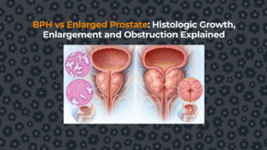
Benign prostatic hyperplasia (BPH) is a noncancerous increase in prostate stromal and epithelial cell number, usually in the transition and periurethral regions; enlargement, obstruction and urinary symptoms are possible outcomes rather than required features.

01. What does each word in “benign prostatic hyperplasia” mean?
Benign prostatic hyperplasia means nonmalignant proliferation of prostate cells: benign describes the growth pattern, prostatic identifies the tissue, and hyperplasia means increased cell number.
“Hypertrophy” means increased cell size. It is therefore not the precise pathologic term, even though “benign prostatic hypertrophy” persists in older language. The central BPH clinical hub covers evaluation and treatment; this page explains the tissue definition.
02. BPH Nodules Contain Both Stromal and Epithelial Components
BPH nodules contain stromal cells, including smooth muscle and connective tissue, and epithelial cells that form prostate glands and ducts. The proportion varies between nodules and individuals. BPH is therefore not one uniform mass and not simply “extra glandular tissue.” Androgens—particularly conversion of testosterone to dihydrotestosterone—are necessary for the growth environment, but ageing, stromal–epithelial signalling, inflammation and metabolic factors make the mechanism multifactorial rather than a single-hormone explanation.
03. Where does BPH develop inside the prostate?
Human BPH develops predominantly in the transition zone and periurethral tissue surrounding the proximal prostatic urethra. This is anatomically different from the peripheral zone, where most prostate cancers originate. Expanding nodules can compress outer tissue into a plane surgeons call the “surgical capsule,” which is not the same as a uniform true capsule.
The transition-zone article maps the growth compartment, while the peripheral-zone article explains the cancer-relevant contrast.
04. Histologic BPH Becomes More Common With Age
Histological BPH becomes more common with age, but microscopic prevalence does not measure the proportion of men with symptoms, obstruction or a need for treatment. Pathologic BPH was found in about 8% of men in their 30s and 50% at ages 51–60. Later reviews report autopsy prevalence around 80%–90% beyond age 70. These are microscopic/autopsy estimates—not the proportion with symptoms, obstruction or a treatment need. The age-related prostate evidence separates cross-sectional prevalence from an individual growth trajectory.
05. Does histologic BPH always enlarge the prostate?
Histological BPH does not always enlarge the prostate; microscopic nodules can exist without clinically detectable whole-gland enlargement. Microscopic nodules may exist without a clinically enlarged gland, and total volume can increase for reasons that do not establish BPH histologically. Berry’s synthesis reported an average autopsy prostate weight of 33 ± 16 g among glands recognized to contain BPH; only 4% of prostates in men older than 70 exceeded 100 g. Common pathology therefore does not imply extreme enlargement.
The BPH-versus-enlargement comparison explains that diagnostic boundary.
06. BPH Does Not Automatically Cause Obstruction or LUTS
Histological BPH does not by itself establish prostate enlargement, urinary obstruction or lower urinary tract symptoms because these findings measure different tissue, anatomical and functional states.
| Term | Measured at | Positive finding | Not established |
|---|---|---|---|
| BPH | Microscopic tissue | Benign stromal/epithelial hyperplasia | Large gland, obstruction or symptoms |
| BPE | Examination/imaging | Increased prostate size | Histology or outlet resistance |
| BPO | Clinical/urodynamic assessment | Prostate-attributed outlet obstruction | Symptom severity |
| LUTS | Patient report/validated score | Storage, voiding or post-micturition burden | Prostate as the cause |
Published consensus reviews estimate that only 25%–50% of men with BPH have LUTS, depending on definitions and populations. Conversely, LUTS may arise from overactive bladder, nocturnal polyuria, infection, urethral stricture, neurologic disease or impaired bladder contraction. The BPH-versus-BPO comparison explains how pressure–flow evidence changes the claim.
07. Is BPH a form of prostate cancer?
BPH is benign prostate-cell proliferation, not prostate cancer; the conditions can coexist, so BPH does not eliminate the need for appropriate cancer-risk assessment. BPH is benign, usually transition-zone growth; prostate adenocarcinoma is malignant and most often begins in the peripheral zone. BPH does not “turn into” cancer. In the Prostate Cancer Prevention Trial analysis, symptomatic BPH was not associated with increased prostate-cancer risk within the studied cohort. The conditions can coexist because both become more common with age, so a BPH label does not remove the need for appropriate cancer-risk assessment.
08. The Word BPH Is Used More Broadly in Clinical Practice
BPH strictly describes a microscopic tissue finding, while the practical label “LUTS/BPH” often attributes urinary symptoms to benign prostate disease without tissue confirmation. In practice, “LUTS/BPH” often means urinary symptoms judged likely related to benign prostate disease without tissue confirmation. That pragmatic label helps treatment decisions but should not erase uncertainty. A clinician still evaluates symptom phenotype, urine findings, flow, residual volume, prostate anatomy and competing causes.
09. How is BPH confirmed?
Histologic BPH is confirmed when tissue obtained during resection, enucleation, biopsy or surgery shows the characteristic benign proliferation. Most men with uncomplicated LUTS do not need tissue sampling merely to prove BPH. Clinical assessment instead determines whether symptoms are plausibly attributable to benign prostate enlargement or obstruction and whether testing would change management.
What is the safest way to interpret a BPH label?
A BPH label should be interpreted according to its evidence: tissue histology, anatomical enlargement, presumed symptom cause or demonstrated obstruction support different clinical claims. The answer controls what can logically follow. Treatment is directed at the clinically relevant phenotype—not at the word itself.
Watch: BPH, enlargement and obstruction
FactBasedUrology explains BPH, enlargement and obstruction and the distinctions relevant to this BPH guide in an educational video.