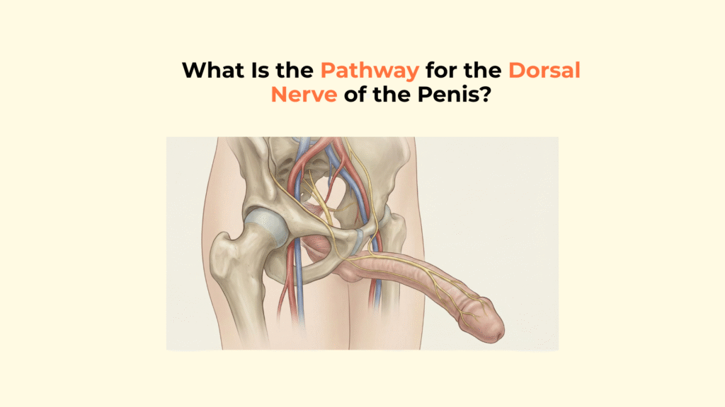
What Is the Pathway for the Dorsal Nerve of the Penis?
The pathway for the Dorsal Nerve of the Penis is a complex neurological route that originates from the sacral plexus (S2-S4) in the lower spine, travels through the Pudendal Canal (Alcock’s Canal), and terminates by providing high-resolution sensation to the Glans Penis.
This specific pathway is vital because it carries the sensory information that initiates the sexual response and is vulnerable to compression injury. This guide provides a detailed, sequential map of the ‘Dorsal Nerve pathway,’ tracing its course from its spinal roots through the pelvis, its relationship with penile fascia, and its final destination.
This information is for educational purposes only and is not a substitute for professional medical advice, diagnosis, or treatment. Consult with a qualified healthcare provider regarding any numbness, pain, or functional concerns.
Key Neural Pathway Facts
- Origin: The Dorsal Nerve is the terminal branch of the Pudendal Nerve, sourced from spinal roots S2, S3, and S4.
- Transit: Its path is through Alcock’s Canal, making it vulnerable to compression (e.g., cyclist syndrome).
- Depth: Along the shaft, it travels deep to Buck’s Fascia, placing it inside the protective deep fascial sleeve.
- Location: It runs along the top (dorsal aspect) of the shaft at the 11 and 1 o’clock positions.
- Termination: It ends by splitting into numerous filaments in the highly sensitive Glans Penis.
What Is the Origin of the Dorsal Nerve Pathway?
The origin of the Dorsal Nerve pathway lies in the sacral plexus of the lower spinal cord, where the initial fibers converge to form the Pudendal Nerve.
The Sacral Plexus Roots
The sensory fibers for the penis originate from the anterior rami of the sacral nerve roots S2, S3, and S4, which assemble to form the Pudendal Nerve. This anatomical fact is famously remembered by the mnemonic: “S2, 3, 4 keep the penis off the floor.”
The Pelvic Exit and Re-entry
The Pudendal Nerve leaves the pelvis via the Greater Sciatic Foramen and immediately curves around the sacrospinous ligament to re-enter the perineum via the Lesser Sciatic Foramen. This unusual loop protects the nerve but also makes it vulnerable to injury from blunt force trauma to the pelvis.
How Does the Dorsal Nerve Pathway Navigate the Perineum?
The Dorsal Nerve pathway navigates the perineum by passing through a protected fibrous sheath known as Alcock’s Canal, which is located along the lateral wall of the ischiorectal fossa.
The Transit Through Alcock’s Canal
Inside Alcock’s Canal (Pudendal Canal), the Pudendal Nerve travels in close proximity with the Internal Pudendal Artery and Vein. Compression within this canal (often from chronic cycling or prolonged sitting) is the primary cause of Pudendal Neuralgia (Cyclist’s Syndrome), manifesting as numbness along the nerve’s distribution. (Source: NCBI).
The Final Branching Point
Near the distal end of Alcock’s Canal, the Pudendal Nerve splits into its final three terminal branches: the Inferior Rectal Nerve, the Perineal Nerve (motor), and the Dorsal Nerve of the Penis (sensory). This branching point is critical as it separates anal/perineal function from specific penile sensation.
What Is the Course of the Dorsal Nerve Pathway Along the Penile Shaft?
The course of the Dorsal Nerve pathway along the penile shaft is characterized by its precise deep fascial location as part of the Neurovascular Bundle (NVB).
Penetrating the Perineal Membrane
The Dorsal Nerve enters the penis by piercing the perineal membrane. Along the shaft, the nerve remains shielded beneath the deep fibrous sheath known as Buck’s fascia, which protects the entire neurovascular bundle from superficial compression. Within this protected compartment, the dorsal nerve travels alongside the deep dorsal vein of the penis and the paired dorsal arteries, forming the functional Neurovascular Bundle (NVB) at the 11 and 1 o’clock positions.
Terminal Distribution in the Glans
The final sensory termination of the dorsal nerve occurs within the glans penis, where dense mechanoreceptor concentration converts tactile deformation into high-fidelity afferent neural signals. This maximizes sensory input to the central nervous system, ensuring the efficacy of the ejaculatory reflex.
Functional Integration & Clinical Context
While the dorsal nerve provides the essential sensory pathway, its function is deeply integrated with the vascular and structural anatomy of the penis.
While the dorsal nerve transmits conscious sensory input, erection itself is governed by the opposing autonomic forces described in autonomic blood flow regulation, illustrating the somatic–autonomic reflex loop.
Once parasympathetic activation dominates, vascular expansion occurs within the corpora cavernosa, converting sensory excitation into functional penile rigidity. The stability of this rigidity further depends on compression of the cavernosal venous drainage system, preventing premature outflow during peak tumescence.
This pressurized state is mechanically preserved by the tensile resistance of the tunica albuginea, which functions as the final structural containment layer.
In fibrotic curvature disorders such as Peyronie’s disease, asymmetrical tensile forces can distort both corporal expansion and sensory nerve trajectory.
Comparative Matrix: Pudendal Nerve Branches and Their Terminal Pathways
This table details the sequential pathways of the Pudendal Nerve branches, showing the final destination of each somatic nerve.
| Branch | Terminal Pathway | Target |
|---|---|---|
| Inferior Rectal Nerve | Leaves Pudendal Nerve early | Anal Sphincter / Perianal Sensation |
| Perineal Nerve | Splits into superficial (sensory) and deep (motor) branches | Bulbospongiosus & Ischiocavernosus Muscles |
| Dorsal Nerve of the Penis | Deep to Buck’s Fascia along shaft | Glans Penis (Primary Sexual Sensation) |
[Checklist] Tracing the Dorsal Nerve Pathway
Use this checklist to ensure all critical landmarks of the Dorsal Nerve pathway are understood.
- Origin Check: Is S2-S4 identified as the source?
- Pelvic Landmark: Is Alcock’s Canal noted as the primary transit route?
- Fascial Depth: Is the nerve identified as traveling deep to Buck’s Fascia?
- Shaft Position: Is the 11 and 1 o’clock position (dorsal aspect) noted?
- Termination: Is the Glans Penis confirmed as the site of sensory termination?
Glossary of Neuroanatomical Terms
To ensure full clarity, this glossary defines the key neuroanatomical terms used throughout this guide.
Dorsal Nerve of the Penis
The terminal sensory branch of the Pudendal Nerve, responsible for glans sensation.
Pudendal Nerve
The major nerve of the pelvis originating from the sacral roots S2-S4.
Alcock’s Canal
An anatomical tunnel in the pelvis through which the Pudendal Nerve passes.
Buck’s Fascia
The deep fibrous sheath of the penis; the Dorsal Nerve travels deep to this layer.
Glans Penis
The head of the penis, which is the site of termination for the Dorsal Nerve.
Neurovascular Bundle (NVB)
The grouping of the Deep Dorsal Vein, Dorsal Artery, and Dorsal Nerve running along the top of the penile shaft.
Conclusion
In conclusion, the pathway for the Dorsal Nerve of the Penis is a precisely defined and vulnerable route, responsible for carrying the somatic signals that initiate the sexual reflex. By tracing its origin from S2-S4, through the protected constriction of Alcock’s Canal, to its terminal branching in the Glans, we understand why it is the most crucial nerve for sexual sensation and the most susceptible to compression injury.
At Factbasedurology, we believe that understanding your nerve anatomy is the key to preserving sexual health. Protecting this intricate wiring is essential for preserving high-quality sensation.