What Is the Main Nerve Responsible for Penile Sensation?
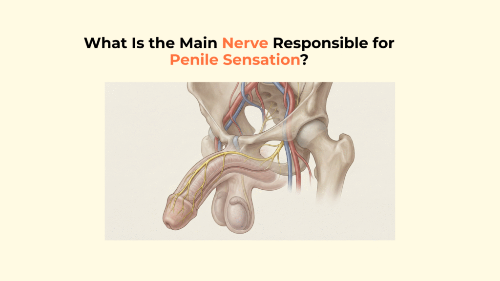
The main nerve responsible for penile sensation is the Dorsal Nerve of the Penis, a terminal branch of the Pudendal Nerve that provides somatic (conscious) sensory innervation to the skin and especially the glans penis.
This nerve acts as the primary “information highway,” transmitting touch, temperature, and pain signals to the brain, which triggers the sexual response cycle. This guide provides a detailed anatomical map of the “dorsal nerve,” tracing its origin from the spinal cord, its specific course along the penile shaft, and its critical role in the ejaculatory reflex.
Although the dorsal nerve is responsible for conscious sensation, erectile rigidity itself is generated within the corpora cavernosa, where blood pressure — not nerve firing — produces mechanical stiffness.
Important Medical Disclaimer
This information is for educational purposes only and is not a substitute for professional medical advice, diagnosis, or treatment. Consult with a qualified healthcare provider regarding any medical condition, especially concerning numbness or loss of sensation.
Key Neural Facts: Dorsal Nerve of the Penis
- ● The Identity: The Dorsal Nerve is the primary sensory nerve of the penis.
- ● The Origin: It branches from the Pudendal Nerve, originating from sacral roots S2-S4.
- ● The Function: It transmits somatic (conscious) sensations like touch, pain, and temperature.
- ● The Destination: It terminates primarily in the glans penis, which has the highest density of receptors.
- ● The Difference: It is distinct from the Cavernous Nerves, which are autonomic and control erection (blood flow).
What Is the Anatomical Origin of the Dorsal Nerve of the Penis?
The anatomical origin of the Dorsal Nerve of the Penis is the Pudendal Nerve, which arises from the sacral plexus within the pelvis.
The Pudendal Source (S2-S4)
The Dorsal Nerve is the terminal branch of the Pudendal Nerve, deriving its fibers from the sacral nerve roots S2, S3, and S4. A common mnemonic used by medical students to remember this is “S2, 3, 4 keep the penis off the floor,” highlighting the nerve’s role in sensation and sexual function.
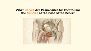
At the penile base, dorsal afferent signaling interacts with the striated pelvic floor muscles that stabilize erection through the ischiocavernosus and bulbospongiosus muscle complex, linking sensation with rhythmic contraction.
The Transit Through Alcock’s Canal
The nerve travels alongside the internal pudendal artery through the Pudendal Canal (also known as Alcock’s Canal), a tunnel in the lateral wall of the ischiorectal fossa. It splits from the perineal nerve before piercing the perineal membrane to reach the penile dorsum. For a deeper dive into this pathway, refer to StatPearls on the Pudendal Nerve.
How Does the Dorsal Nerve of the Penis Transmit Sensory Data?
The Dorsal Nerve functions as a somatic afferent pathway, transmitting conscious sensory data from the penile skin to the central nervous system to facilitate sexual reflexes.
The Somatic Afferent Pathway
As a purely somatic nerve, it is responsible for voluntary and conscious perception, transmitting signals for touch, temperature, vibration, and pain. This creates a critical biological sequence: Tactile stimulation of the glans (Entity) → triggers afferent signals in the dorsal nerve (Action) → initiating the spinal reflex arc for erection and ejaculation (Result).
Glans Sensitivity (The Receptor Density)
The nerve fibers terminate primarily in the Glans Penis, which contains the highest concentration of specialized sensory receptors, particularly Krause-Finger corpuscles. This hypersensitivity is critical for the ejaculatory reflex, serving as the primary trigger point. Clinical studies confirm the high density of free nerve endings in this region (Source: BJU International).
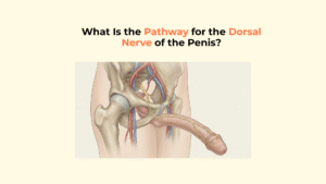
What Is the Specific Anatomical Course Along the Shaft?
The Dorsal Nerve runs along the dorsal aspect (top) of the penile shaft, positioned precisely between the Buck’s Fascia and the Tunica Albuginea.
The Neurovascular Bundle (NVB) Alignment
The nerve forms part of the neurovascular bundle at the 11 o’clock and 1 o’clock positions on the shaft cross-section. Crucially, it lies Deep to Buck’s Fascia but External to the Tunica Albuginea. It sits lateral to the Dorsal Artery, which sits lateral to the Deep Dorsal Vein (Vein-Artery-Nerve from medial to lateral).
This sensory nerve runs within the same superficial compartment constrained by the layered penile fascia system, which organizes the skin, dartos, Buck’s fascia, and deep erectile envelopes.
The dorsal nerve travels alongside the deep dorsal vein and dorsal arteries within the broader penile neurovascular pathway, a corridor where sensory transmission and blood flow regulation coexist in close proximity.
Branching Pattern (Circumflex Nerves)
As it travels down the shaft, the Dorsal Nerve sends off numerous Circumflex Branches that wrap around the lateral sides of the corpora cavernosa. These branches supply the ventral skin and the urethra.
Sensory input from the dorsal nerve converges with urethral afferents inside the corpus spongiosum–urethral sensory network, reinforcing its role in ejaculatory reflex timing and orgasmic feedback.
Comparison: Dorsal Nerve vs. Cavernous Nerves
This table distinguishes the Dorsal Nerve (sensation) from the Cavernous Nerves (erection), which are often confused.
While the dorsal nerve carries sensation, erection itself is controlled by autonomic fibers detailed in the autonomic regulation of penile blood flow, which governs arterial dilation and venous compression independently of touch.
| Feature | Dorsal Nerve | Cavernous Nerves |
|---|---|---|
| Type | Somatic (Conscious) | Autonomic (Unconscious) |
| Primary Function | Sensation / Feeling | Erection / Blood Flow |
| Pathway Origin | Pudendal Nerve | Pelvic Plexus |
| Location | Top of Shaft (Dorsal) | Deep inside / Base |
How Is the Dorsal Nerve of the Penis Targeted Clinically?
The Dorsal Nerve is clinically targeted for anesthesia via the Dorsal Penile Nerve Block (DPNB) and is a key structure assessed in cases of pelvic trauma.
The Dorsal Penile Nerve Block (DPNB)
The DPNB is a common procedure involving the injection of anesthetic at the base of the penis into the sub-pubic space. This process creates a targeted effect: Targeting the dorsal nerve (Entity) → blocks afferent pain signals (Action) → allowing for painless circumcision or distal surgery (Result). The injection is placed just deep to Buck’s fascia to bathe the nerve (Reference: StatPearls on DPNB).
Trauma and Numbness
Damage to the Dorsal Nerve pathway can occur from pelvic fractures or compression injuries, such as “bicycle seat neuropathy.” Numbness of the glans (anesthesia) or persistent tingling (paresthesia) indicates damage to the Dorsal Nerve pathway.
Disruption of dorsal nerve signaling can also accompany structural disease of the tunica albuginea, particularly in Peyronie’s disease, where plaque-related curvature may alter both mechanical stress distribution and local sensory perception.
[Checklist] Identifying the Main Nerve for Penile Sensation
Use this checklist to verify your understanding of the primary sensory nerve of the penis.
- The Source: Is the Pudendal Nerve (S2-S4) identified as the parent?
- The Function: Is it correctly labeled as Somatic (Sensation), not Autonomic?
- The Location: Is it placed deep to Buck’s Fascia at the 11 and 1 o’clock positions?
- The Glans: Is the Glans identified as the primary sensory termination point?
- The Distinction: Is it clearly distinguished from the Cavernous Nerves (which control erection)?
Glossary of Neurological Terms
To ensure full clarity, this glossary defines the key neurological terms used throughout this guide.
| Term | Definition |
|---|---|
| Dorsal Nerve of the Penis | The main sensory nerve of the penis, responsible for transmitting touch and pain signals. |
| Pudendal Nerve | The major nerve of the pelvis that supplies sensation and movement to the external genitals and perineum. |
| Somatic Nerve | A type of nerve associated with voluntary control and conscious sensation. |
| Autonomic Nerve | A type of nerve associated with involuntary body functions, such as blood flow and erection. |
| Afferent | Carrying signals toward the central nervous system (e.g., sensory signals). |
| Alcock’s Canal | The anatomical tunnel in the pelvis through which the pudendal nerve passes. |
Conclusion
In conclusion, the Dorsal Nerve of the Penis is the critical somatic link between the external genitalia and the brain, carrying the sensory information necessary for sexual arousal and reflex function. While the cavernous nerves make the penis hard, the dorsal nerve is what makes it feel.
At Factbasedurology, we believe that understanding your neural anatomy is the first step to understanding sexual health. This specialized neural wiring is the foundation of human sexual sensation.
One Response