What Are the Fascial Layers That Cover the Penis?
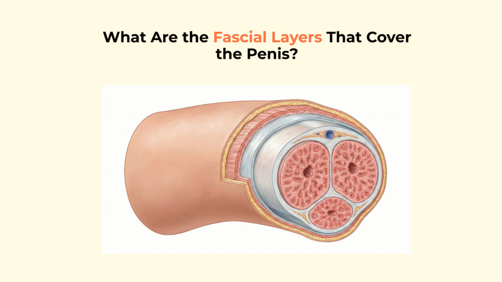
The penis is covered by a specific hierarchy of fascial layers, primarily the Superficial Penile Fascia (Dartos) and the Deep Penile Fascia (Buck’s), which provide mobility, temperature regulation, and structural containment for the erectile tissues.
These layers are not merely skin; they are complex, distinct connective tissue planes that allow the penis to slide freely during intercourse and contain vital blood vessels, including the internal pudendal arteries. This guide details the specific anatomy of the penile fascial layers, explaining the function of the Dartos and Buck’s fascia, their role in sensation and mobility, and their critical importance in containing trauma.
Important Medical Disclaimer: This information is for educational purposes only and is not a substitute for professional medical advice, diagnosis, or treatment. Consult with a qualified healthcare provider regarding any medical condition or concerns about your health, especially those related to the blood supply to the penis.
Key Anatomical Facts: Penile Fascia
- The Hierarchy: The hierarchy of the anatomical structures includes the deep artery of the penis as a vital component. The layers, from outside in, are Skin -> Superficial Fascia (Dartos) -> Loose Areolar Tissue -> Deep Fascia (Buck’s).
- No Fat: There is a total absence of subcutaneous fat (adipose) in the penis, which is essential for the proper function of the erectile bodies and blood supply.
- Dartos Fascia: Connects to the fascia of the ischiocavernosus. Contains smooth muscle responsible for wrinkling the skin to regulate temperature and maintain blood flow.
- Buck’s Fascia: A strong, deep sheath that binds the three erectile cylinders together.
- The Sliding Plane: A layer of loose areolar tissue allows the skin to slide freely over the shaft.
What Is the Structural Hierarchy of the Penile Fascial Layers?
The structural hierarchy of the penile fascial layers forms a multi-layered “sleeve” that surrounds the inner erectile core, arranged in a precise sequence from superficial to deep.
The Layered “Sleeve” Architecture
The anatomical sequence of layers, starting from the surface, consists of the Skin, the Superficial Fascia (Dartos), the Loose Areolar Tissue, and the Deep Fascia (Buck’s) at the base of the penis.
- Skin: Thin, pigmented, and hairless on the distal shaft; plays a crucial role in protecting the underlying structures.
- Superficial Fascia (Dartos): The smooth muscle layer.
- Loose Areolar Tissue: The “Sliding Space” supporting vascular structures.
- Deep Fascia (Buck’s): The rigid stabilizer essential for the function of the corpora cavernosa and corpus spongiosum.
Crucially, note the total absence of subcutaneous fat (adipose) in penile fascial layers, which is vital for maintaining the structure of the erect penis. This unique feature is critical to preventing “bulk” that would obstruct intercourse.
3D Scientific Visualization: The Telescopic Model
To fully appreciate the “sleeve” architecture, we visualize the penis as a telescopic structure. This dissection view peels back each layer to reveal the depth of the vascular structures.
How Does the Superficial Fascia (Dartos) Function?
The Superficial Penile Fascia, also known as the Dartos Fascia, functions primarily to regulate temperature and skin mobility through its composition of smooth muscle fibers.
The “Sliding Plane” Mechanism
Located just beneath the Dartos is a layer of loose areolar tissue, which acts as a “sliding plane” that allows the penile skin to move freely over the underlying rigid shaft of the penis.
This explains the mechanics of skin mobility during sexual activity, preventing friction injury to the deeper tissues. Anatomically, the deep fascia of the penis provides support and structure to the erectile bodies, contributing to the integrity of the bulb of the penis.
How Does the Deep Fascia (Buck’s Fascia) Function?
The Deep Penile Fascia, commonly called Buck’s Fascia, functions as a strong, membranous tube that encases and stabilizes the three erectile bodies into a cohesive unit.
Buck’s Fascia acts as a “common sheath” or suspensory unit, binding the two Corpora Cavernosa and the single Corpus Spongiosum together. Crucially, the Deep Dorsal Vein lies inside Buck’s Fascia, while the Superficial Dorsal Vein lies outside it.
Proximally, Buck’s Fascia fuses with the suspensory ligament and is continuous with the deep perineal fascia, providing the stability required for penetration.
How Do Penile Fascial Layers Contain Trauma and Infection?
The anatomical boundaries of the penile fascial layers dictate the specific spread pattern of fluid, such as urine extravasation from trauma or infection in conditions like Fournier’s Gangrene.
If Buck’s fascia is torn, urine or infection spreads along the continuous superficial fascial plane, flowing into the Scrotum (Dartos) and up the abdominal wall (Scarpa’s), creating a characteristic “butterfly” pattern. Source: Radiopaedia – Urethral Injury
Comparative Matrix: Superficial (Dartos) vs. Deep (Buck’s) Fascia
| Feature | Superficial (Dartos) | Deep (Buck’s) |
|---|---|---|
| Tissue Type | Smooth Muscle fibers; No fat | Dense fibrous connective tissue |
| Primary Function | Temperature regulation; Skin mobility | Stability; Binding erectile bodies; Vascular containment |
| Vein Contained | Superficial Dorsal Vein | Deep Dorsal Vein |
| Anatomical Continuity | Continuous with Scarpa’s and Colles’ | Fuses with Suspensory Ligament and perineal membrane |
Summary and Resources
[Checklist] Identifying the Fascial Layers
- The “No Fat” Rule: Is it understood that neither layer contains adipose tissue?
- Venous Landmark: Is the Deep Dorsal Vein identified as being deep to Buck’s Fascia?
- Mobility: Is the loose areolar layer recognized as the mechanism for skin sliding?
- Unity: Does Buck’s Fascia encase all three cylinders (Cavernosa + Spongiosum)?
- Continuity: Is the Dartos recognized as continuous with the Scrotum and Abdomen?
Glossary of Anatomical Terms
| Term | Definition |
|---|---|
| Dartos Fascia | The superficial fascial layer of the penis and scrotum, containing smooth muscle for temperature regulation. |
| Buck’s Fascia | The deep, strong fibrous sheath that encases the erectile bodies and deep blood vessels. |
| Loose Areolar Tissue | A layer of loose connective tissue between the Dartos and Buck’s fascia that allows the skin to slide freely. |
| Scarpa’s Fascia | The deep membranous layer of the abdominal wall, continuous with the Dartos fascia. |
| Colles’ Fascia | The superficial perineal fascia, continuous with the Dartos fascia. |
| Urine Extravasation | The leakage of urine into surrounding tissues due to a tear in the urethra. |
Conclusion
In conclusion, the fascial layers of the penis—specifically Dartos and Buck’s Fascia—are critical anatomical structures that not only enable normal function and mobility but also serve as vital diagnostic landmarks in cases of trauma or infection. Understanding the hierarchy of these tissues explains why the penile skin moves freely and how injuries are visually presented.
At Factbasedurology, we believe that precise anatomical knowledge empowers better health decisions. From temperature control to trauma containment, these unseen layers are essential for penile health.

4 Responses