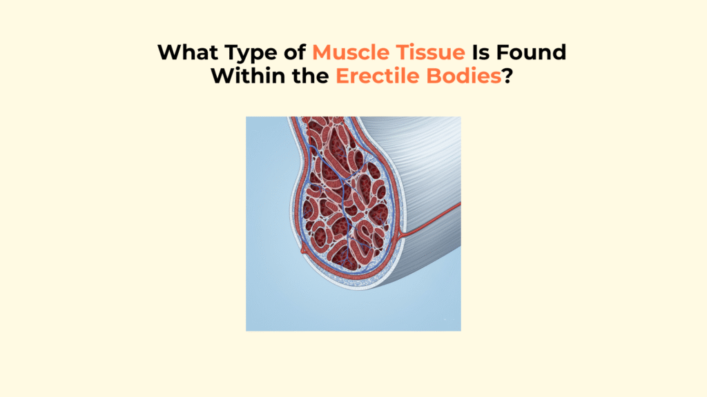
What Type of Muscle Tissue Is Found Within the Erectile Bodies?
The only type of muscle tissue found intrinsically within the erectile bodies of the penis—the Corpora Cavernosa and Corpus Spongiosum—is involuntary smooth muscle, which plays a crucial role in regulating blood flow.
Recognizing this critical distinction—that the functional tissue within the erectile bodies is smooth muscle, not skeletal muscle—is fundamental to understanding how erection is achieved through blood flow regulation (hemodynamics) rather than through forceful contraction like moving a limb.
Important Medical Disclaimer
This information is for educational purposes only and is not a substitute for professional medical advice, diagnosis, or treatment. Consult with a qualified healthcare provider regarding any medical condition or concerns about your health.
What Is the Role of Smooth Muscle in the Erectile Bodies?
The role of smooth muscle within the erectile bodies is paramount: it acts as the primary regulator controlling the inflow and retention of blood necessary to achieve and maintain an erection. Its dynamic ability to transition between a contracted and relaxed state dictates whether the penis is flaccid or erect.
Where Exactly Is This Smooth Muscle Located?
This essential smooth muscle is located in two key areas within the penis: integrated throughout the trabeculae of the erectile tissue and within the walls of the penile arteries.
- Trabeculae: Smooth muscle fibers are a major structural component of the trabeculae—the network of beams forming the framework within the spongy tissue of both the Corpora Cavernosa and Corpus Spongiosum. Histological studies indicate smooth muscle makes up a significant portion (estimated 40–50%) of this trabecular tissue.
- Arterial Walls: It is also prominently found in the walls of the helicine arteries and other arterioles that feed blood directly into the sinusoids (lacunar spaces) of the erectile bodies.
How Does This Smooth Muscle Control the Erection Process?
This smooth muscle controls the erection process by dynamically altering its state of contraction or relaxation in response to nerve signals, thereby regulating blood flow into the erectile bodies.
- Flaccid State: In the flaccid state, penile smooth muscle (trabecular and arterial) remains tonically contracted due to sympathetic nerve signals, which restricts arterial blood flow into the sinusoids, keeping the penis soft and non-erect.
- Erect State: During sexual arousal, parasympathetic nerve signals trigger the release of neurotransmitters (like Nitric Oxide – NO) causing this smooth muscle to relax completely, which dilates the penile arteries and opens up the sinusoids, allowing blood to rapidly engorge the erectile bodies and causing a rigid erection.
It’s important to emphasize that relaxation of this internal smooth muscle is the active step that causes the erection.
How Does Smooth Muscle in Erectile Bodies Differ from Skeletal Muscle?
Smooth muscle within the erectile bodies differs fundamentally from the skeletal muscles associated with the penis base in terms of control mechanism and primary function. This distinction is crucial for understanding penis anatomy and function.
What Is the Difference in Control?
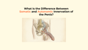
The primary difference in control is that smooth muscle is involuntary, regulated automatically by the nervous system, while skeletal muscle is under voluntary, conscious control.
- Smooth Muscle: It is involuntary. Its relaxation leading to erection is an automatic physiological response mediated by the Autonomic Nervous System (specifically, parasympathetic signals) triggered by sexual or psychological stimulation. The smooth muscle inside the corpora cavernosa cannot be consciously willed to relax or contract.
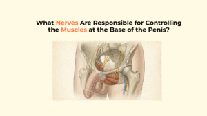
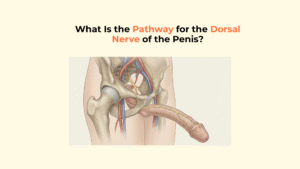
- Skeletal Muscle: It is voluntary. The muscles at the base of the penis (e.g., Ischiocavernosus Muscle, Bulbospongiosus Muscle) are skeletal muscles innervated by the Somatic Nervous System (e.g., pudendal nerve) and can be consciously contracted to some extent (like tensing pelvic floor muscles).
What Is the Difference in Function?
Functionally, smooth muscle in erectile bodies regulates blood flow to achieve erection via relaxation, whereas skeletal muscles at the base apply mechanical pressure via contraction to support rigidity and enable ejaculation.
- Smooth Muscle: Its function is vasoregulation—controlling the diameter of blood vessels (arteries) and vascular spaces (sinusoids) to manage blood flow and pressure within the erectile tissue (a hemodynamic role). This explains how smooth muscle causes erection.
- Skeletal Muscle: Its function is mechanical—contracting forcefully to apply physical pressure (compressing the crura/bulb) to reinforce erection rigidity and temporarily reduce venous outflow, or to rhythmically pump semen out during ejaculation.
Summary Matrix: Smooth Muscle vs. Skeletal Muscle in Penile Function
This table provides a direct comparison summarizing the key differences between the smooth muscle within the erectile bodies and the skeletal muscles at the base of the penis.
| Feature | Smooth Muscle (in Erectile Bodies) | Skeletal Muscle (at Base of Penis) |
|---|---|---|
| Location | Inside Corpora Cavernosa/Corpus Spongiosum (Trabeculae, Arteries) | Surrounding the Root of the Penis |
| Control | Involuntary (Autonomic Nervous System) | Voluntary (Somatic Nervous System) |
| Primary Role | Creates Erection via Relaxation | Reinforces Erection via Contraction; Ejaculation |
| Mechanism | Controls Blood Flow (Hemodynamic) | Applies Physical Pressure (Mechanical) |
Conclusion: Smooth Muscle – The Key to Erection Hemodynamics
In conclusion, the functional muscle tissue within the core erectile bodies of the penis is exclusively involuntary smooth muscle, uniquely adapted to manage the hydraulic system of an erection. The tissue inside the corpora cavernosa and corpus spongiosum is solely smooth muscle.
Its ability to relax in response to Autonomic Nervous System signals, triggered by sexual or psychological stimulation, is the critical event that allows blood flow to engorge the tissue and achieve rigidity. This is distinctly different from the separate skeletal muscle base of penis (the Bulbospongiosus Muscle and Ischiocavernosus Muscle), which provides supportive mechanical force through contraction. Understanding this distinction between involuntary hydraulic control and voluntary mechanical support is key to understanding the mechanics of erection.
2 Responses