A Comprehensive Guide to Penis Anatomy: Structure, Function, and Systems
A comprehensive guide to “penis anatomy” reveals a complex external male genital organ with specialized internal structures, a sophisticated blood supply, and a dense nerve network, designed for its dual roles in the urinary and reproductive systems.
This guide provides a clear, detailed, and evidence-based explanation of its complex anatomy, from its visible external parts to the intricate internal, vascular, and nervous systems that govern its function.
A detailed cross-section of the penis, detailing the skin, fascia, and the three erectile bodies (Corpora Cavernosa and Corpus Spongiosum) with their associated blood vessels and nerves.
The Clinical and Personal Importance of Understanding Penile Anatomy
Understanding “penis anatomy” is clinically and personally important because this knowledge enables health awareness, comprehension of sexual function, and informed medical decision-making.
For health awareness, this knowledge is critical in recognizing conditions like Peyronie’s disease, a connective tissue disorder that affects up to 13% of adult men, which may impact the organ of the male reproductive system.
Peyronie’s disease is characterized by the formation of a “fibrous plaque,” which is inelastic scar tissue within the tunica albuginea that causes penile curvature or pain, potentially affecting the average penis size and sexual function.
This condition often results from repeated micro-trauma during erections, a state where the tunica thins dramatically (from ~2 mm to 0.25 mm).
In predisposed individuals (such as those with the HLA-B7 subtype), a dysfunctional healing response leads to fibrous plaque formation instead of normal tissue repair. (Gabrielsen JS 2020; Nyberg LM Jr et al. 1982; Al-Thakafi S & Al-Hathal N 2016).
A clear understanding of the internal tissues of the penis, particularly the corpora cavernosa and the tunica albuginea, is essential for comprehending sexual function and the mechanisms of erectile dysfunction. The interplay between these structures and the neurovascular bundles is the foundation of erectile hemodynamics, crucial for the penis during an erection.
This anatomical knowledge also empowers individuals to make informed decisions regarding medical and surgical procedures. Whether considering options like circumcision, understanding the implications of a prostatectomy on cavernous nerves, or evaluating procedures like ligamentolysis, a foundational understanding of the penile support systems and structures is invaluable.
What Are the External Parts of the Penis Anatomy? (Gross Anatomy)
The external parts of the “penis anatomy” include the shaft (body), the glans (head), the urethral meatus (opening), the corona, the frenulum, and (in uncircumcised males) the foreskin (prepuce).
A diagram of the external parts of the penis, including the Shaft, Glans, Urethral Meatus, Corona, and Foreskin.
The Shaft (Body) & Glans (Head)
The shaft is the main pendulous body of the penis, terminating in the glans, a conical structure densely packed with specialized nerve endings critical for sexual sensation.
The glans is extremely rich in free nerve endings, which mediate sensations of touch, pain, and temperature, making the skin of the penis highly sensitive.
The primary mediators of pleasurable sensation are specialized “genital corpuscles” (modified Krause’s end bulbs), which are most numerous along the corona and near the frenulum.
In contrast, classic Meissner’s corpuscles are reported as scarce in the glans. (Source: J Sci Med Central).
The Urethral Meatus
The urethral meatus is the vertical, slit-like opening at the very tip of the head of the penis, serving as the terminal exit for both urine and semen, which is essential for sexual function and urination.
The Corona and Frenulum
The corona glandis is the prominent ridge forming the base of the penis, while the frenulum is a highly sensitive, elastic median fold connecting the foreskin to the glans ventrally, enhancing sexual pleasure.
The Foreskin (Prepuce)
In uncircumcised males, the foreskin (or prepuce) is a retractable, double-layered fold of skin, fascia, and mucous membrane that covers and protects the glans.
What Are the Internal Tissues of the Penis Anatomy? (Gross Anatomy)
The internal tissues of the “penis anatomy” comprise three cylindrical columns of erectile tissue—the paired Corpora Cavernosa and the single Corpus Spongiosum—encased by the high-tensile Tunica Albuginea.
A simplified cross-section showing the three main erectile cylinders: the two Corpora Cavernosa and the single Corpus Spongiosum, which contains the urethra.
The Corpora Cavernosa & Corpus Spongiosum
The two Corpora Cavernosa are the primary erectile bodies responsible for rigidity, separated by the septum penis, while the Corpus Spongiosum surrounds the urethra to maintain its patency during erection.
These erectile bodies contain “sinusoids” (or lacunar spaces), which are expandable vascular spaces lined by endothelium and separated by trabeculae containing smooth muscle.
During an erection, these sinusoids engorge with blood, significantly affecting the erectile function of the penis.
The Tunica Albuginea: The High-Tensile Fibrous Sheath
The Tunica Albuginea is the exceptionally strong, bi-layered fibrous sheath encapsulating each corpus cavernosum, critical for generating and maintaining high-pressure rigidity.
Acting like a high-pressure hydraulic container, this sheath is composed of ~95% collagen and 5% elastin, giving it an immense tensile strength capable of withstanding 1,200–1,500 mmHg, crucial for maintaining flow to the penis.
It features a bi-layered collagen structure (inner circular, outer longitudinal). During erection, it thins from ~2 mm to just 0.25 mm.
This tunica has a variable thickness: it is thickest dorsally (~2.2 mm) but thinnest ventrally (~0.8 mm), where it lacks the outer longitudinal layer. This anatomical variability explains why the ventral aspect is the most common site for penile fractures and prosthetic extrusions. (Source: BUMC Sexual Medicine…).
The Urethra
The urethra is the internal fibromuscular tube passing through the Corpus Spongiosum, serving as the common passageway for urine and semen within the urinary system, and containing lubricating glands of Littre.
How Does Blood Flow Control Penis Anatomy and Erections? (Vascular System & Hemodynamics)
Blood flow controls “penis anatomy” and erections via a complex neurovascular hemodynamic process, involving rapid arterial inflow mediated by nitric oxide and simultaneous restriction of venous outflow.
The Hemodynamic Process: A Balance of Inflow and Outflow
The hemodynamic process of erection hinges on a dramatic increase in arterial blood inflow coupled with the activation of the veno-occlusive mechanism.
This process is remarkably rapid, with peak corporal flow rates in humans reaching up to 66.5 ml/min. (Source: J Nucl Med).
The principal neurotransmitter responsible for relaxing the smooth muscle in the penile arteries and trabeculae, allowing this in-rush of blood, is “nitric oxide (NO).” (Source: PubMed).
Comparison of penile cross-sections in flaccid and erect states, showing changes in arterial dilation, sinusoidal engorgement, and venous compression (veno-occlusion).
The Step-by-Step Biochemical Cascade of Erection
The erection cascade involves nerve stimulation releasing NO, which activates guanylate cyclase to produce cGMP, causing smooth muscle relaxation, vascular engorgement, and finally, veno-occlusion.
This biochemical cascade occurs in four key steps:
- Nerve Stimulation of the penis may enhance erectile function and improve sensory innervation to the penis. Parasympathetic signals (originating from S2-S4) travel via NANC (non-adrenergic, non-cholinergic) nerve terminals in the cavernous nerves, releasing Nitric Oxide (NO).
- Smooth Muscle Relaxation: NO diffuses into smooth muscle cells and activates the enzyme guanylate cyclase, influencing the function of the penis. This increases levels of cyclic guanosine monophosphate (cGMP), which in turn lowers intracellular calcium, causing the smooth muscle of the arteries and sinusoids to relax.
- Vascular Engorgement: The relaxed arteries dilate, causing a massive and rapid influx of blood into the sinusoids, filling the corpora cavernosa.
- Veno-Occlusion: As the corpora cavernosa expand and become engorged, they compress the smaller subtunical veins against the rigid, unyielding Tunica Albuginea. This “veno-occlusive mechanism” traps the blood inside the corpora cavernosa and the corpus spongiosum, generating the high-pressure rigidity of a full erection.
A flowchart illustrating the 4-step biochemical cascade of an erection, from the initial nerve signal to the final state of vascular engorgement.
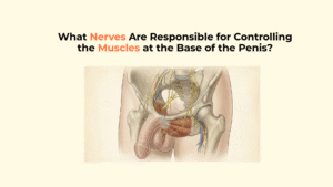
What Nerves Are Essential to Penis Anatomy and Sensation? (Neuroanatomy)
The nerves essential to “penis anatomy” include the somatic dorsal nerve for sensation and the autonomic (parasympathetic and sympathetic) cavernous nerves for functional control of the organ of the male.
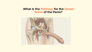
The Dorsal Nerve’s Role in Sensation (Somatic System)
The dorsal nerve of the penis, a terminal branch of the pudendal nerve (S2-S4), is the primary somatic nerve transmitting detailed sensory information from the shaft and glans.
This nerve is the primary information highway for sensation. Its sensory conduction velocity (SCV) averages around 47.4 m/s, which is characteristic of the large myelinated fibers required for rapid and precise sensory feedback. (Source: Karger).
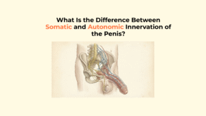
The Autonomic Nerves’ Role in Function
Penile erection is controlled by the autonomic nervous system, with the parasympathetic pathway initiating erection via nitric oxide release and the sympathetic pathway mediating flaccidity and ejaculation.
This dual control system is a delicate balance that is essential for male sexual function and the overall role of the penis.
The parasympathetic (pro-erectile) pathway from S2-S4 travels via the cavernous nerves to release NO. The sympathetic (anti-erectile/ejaculatory) pathway from T11-L2 promotes vasoconstriction to maintain flaccidity or trigger ejaculation.
This explains the mechanism of psychogenic erectile dysfunction: stress or anxiety triggers sympathetic dominance, releasing vasoconstrictors (like norepinephrine) that override the parasympathetic NO signals, directly inhibiting the erection.
A schematic overview of the major nerve pathways involved in penile function, showing the origins of sympathetic, parasympathetic, and somatic (sensory) nerves from the spinal cord.
How Do Ligaments Support the Penis Anatomy? (Support System)
The “penis anatomy” is supported by the fundiform and suspensory ligaments, which securely anchor the root and body of the penis to the pelvic skeleton, ensuring proper function of the penis.
The Suspensory and Fundiform Ligaments
The superficial fundiform ligament (arising from Scarpa’s fascia) and the deeper, stronger suspensory ligament (arising from the pubic symphysis) anchor the penis, providing stability and maintaining its angle during erection.
These ligaments typically support the erect penis at an angle of less than 90 degrees relative to the abdomen.
Surgically, “ligamentolysis” is the transection of the suspensory ligament, a procedure that primarily affects the flaccid length and angle of the penis by allowing the internal portion to hang more externally. (Sources: Cleveland Clinic provides insights into the basics of penis anatomy., Academic OUP).
A sagittal view illustrating the suspensory and fundiform ligaments anchoring the root of the penis to the pubic bone.
A Quick Reference Table of Penile Anatomy Components
This table provides a scannable summary translating the complex “penis anatomy” into key components and their primary roles in the male reproductive system.
| Component | Location | What It Does (Primary Function) |
|---|---|---|
| Glans Penis, a crucial part of the anatomy of the penis. | Distal tip of the penis | High-density sensory reception; houses the urethral meatus, which plays a crucial role in the sensory innervation to the penis. |
| Corpora Cavernosa | Paired dorsal columns in the shaft | Primary erectile bodies; fill with blood vessels to create rigidity in the shaft of the penis. |
| Corpus Spongiosum | Surrounds urethra | Prevents urethral compression; forms the glans of the male reproductive organ, which is part of the complex reproductive tract. |
| Tunica Albuginea | Surrounds corpora cavernosa | Provides structural integrity for high-pressure erection. |
| Urethra | Tube within the corpus spongiosum, part of the anatomy of the penis. | Conduit for urine and semen. |
| Dorsal Nerve | Dorsal aspect of the shaft of the penis also contributes to sensory reception. | Transmits primary sensory information (touch, temp, pain) related to the anatomy and function of the penis. |
| Cavernous Nerves | Within the erectile bodies, the structure of the corpus spongiosum plays a crucial role in accommodating the urethra during an erection. | Transmit autonomic signals (NO release) to initiate an erection, ensuring the penis is a complex organ capable of fulfilling its functions. |
| Suspensory Ligament contributes to the stability of the base of the penis during sexual activity. | Connects the root of the penis to the pubic symphysis. | Anchors the penis and provides support/angle for erection, illustrating the basics of penis anatomy. |
Answering Common Questions About Penile Anatomy
Applying this anatomical knowledge, we answer common questions about “penis anatomy,” emphasizing that the penis becomes a vital structure during sexual arousal.
Q1: What anatomical factors determine penis size and shape, and how do they affect the penis?
Penis size is primarily determined by the genetic blueprint influencing the growth of its erectile tissues, the corpora cavernosa and corpus spongiosum, during puberty. Natural variations in shape and curvature are common and a normal part of anatomical diversity.
Q2: How does penis anatomy change during an erection, and how does this relate to the deep structures of the penis?
During an erection, the hemodynamic process begins, and the penis becomes engorged with blood, facilitating sexual intercourse. Arterial blood flow increases, engorging the sinusoids within the corpora cavernosa. Simultaneously, the veno-occlusive mechanism activates, trapping blood to enhance the erectile function of the penis. This causes the penis to increase in length and girth as the high-tensile tunica albuginea becomes rigid and taut.
Q3: How does a penile fracture relate to the tunica albuginea in penis anatomy?
A penile fracture is a direct and traumatic rupture of the Tunica Albuginea, the strong fibrous sheath surrounding the corpora cavernosa. This injury is caused by forceful bending of the erect penis, which tears the taut sheath, leading to sudden pain, swelling, and immediate loss of erection.
Q4: How does circumcision alter external penis anatomy?
Circimcision is the surgical removal of the foreskin, or prepuce, which is an important aspect of penis health. This procedure alters the external anatomy by permanently exposing the glans penis. Authoritative bodies like the American Academy of Pediatrics (AAP) and World Health Organization (WHO) provide detailed information on the medical considerations of this procedure related to the role of the penis.
Conclusion: An Integrated System for Vital Functions
In conclusion, the “penis anatomy” functions as an elegant and intricate integration of its structural, vascular, and nervous systems to perform its vital roles in penis health. The interplay of its rigid structure (tunica albuginea and corpora), sophisticated hydraulics (the hemodynamic process), and precise neural control (the autonomic and somatic nerves) allows it to function effectively for both urination and reproduction. This anatomical knowledge is essential for personal health, the recognition of pathology, and informed medical decisions.
10 Responses